Introduction
Among the cutaneous extraintestinal manifestations of inflammatory bowel disease (IBD), metastatic Crohn’s disease (MCD) occupies a unique position, being both the rarest and one of the most disfiguring of these entities. It is defined as non-caseating granulomatous inflammation arising in the skin or mucous membranes at sites unconnected with the gastrointestinal tract. Histologically, MCD thus represents the cutaneous counterpart of the lesions seen in Crohn’s disease [1, 2]. Although until recently regarded as a rarity, patients with MCD now require collaboration between gastroenterology, dermatology and, frequently, rheumatology.
A granulomatous disease that crosses specialty boundaries
Metastatic Crohn’s disease was first described in 1965 by Parks [1]. Over the subsequent 6 decades, only about 500 cases have been reported worldwide, most of them solely as single case reports and small case series [2, 3]. It is generally accepted that, owing to low disease awareness among clinicians, the true incidence is markedly underestimated, with many cases remaining unrecognised or misclassified.
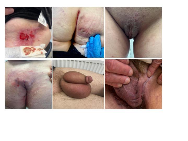
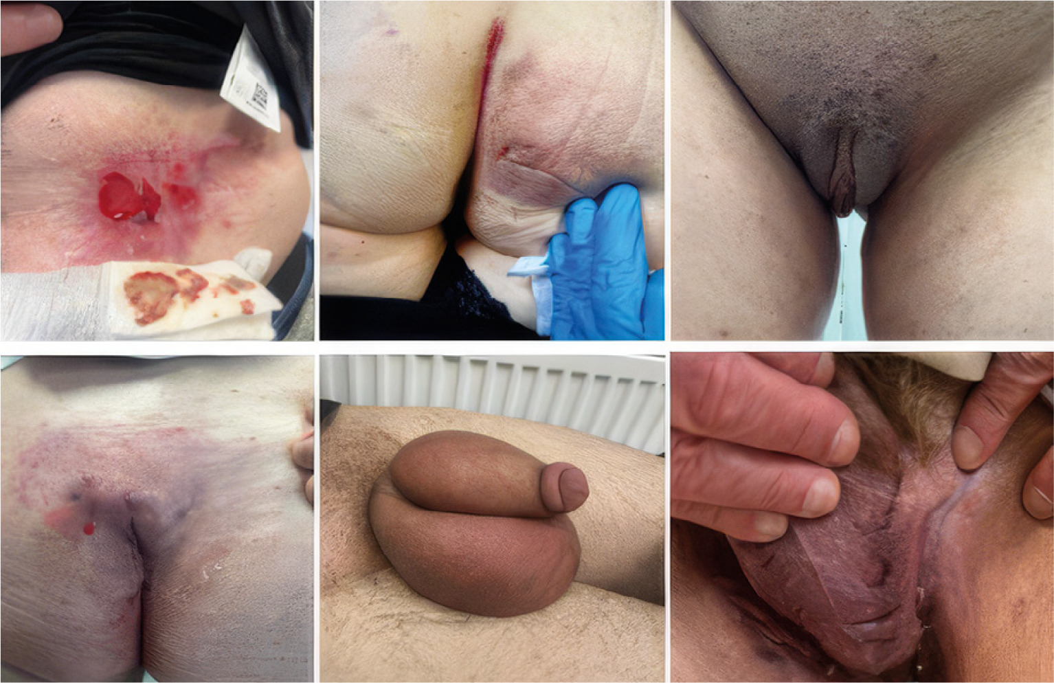
A source of diagnostic difficulty is the fact that MCD shares a diagnostic challenge similar to that of other granulomatous dermatoses. Clinically, the lesions are polymorphic: isolated, chronic swelling of the labia, scrotum or perineum, erythematous-infiltrative changes, and nodules, often with characteristic linear “knife-cut” fissures that are particularly pronounced in the genital and intertriginous skin (Fig. 1). The lesions frequently leave scars or permanent deformity in the anogenital region. Because there is no pathognomonic feature, MCD may be confused with infections, hidradenitis suppurativa, pyoderma gangrenosum, erythema nodosum and – most closely on histology – sarcoidosis and Behçet’s disease [2, 4].
Fig. 1
Heterogeneity of the clinical presentation of metastatic Crohn’s disease across anatomical sites.
The diagnosis is further complicated by the temporal course. Cutaneous lesions most often appear after the diagnosis of intestinal Crohn’s disease, concurrently with it, or precede it; moreover, the skin lesions follow a course entirely distinct from intestinal activity, flaring during remission of bowel disease and persisting despite endoscopic remission [2, 5]. This discordance between cutaneous and intestinal activity is more than a clinical inconvenience – it indicates that the immune response in the skin behaves autonomously, independently of the process in the gut. It is explained by the contemporary model of MCD pathogenesis. Its central effector axis is well recognised in rheumatology: dysregulation of Th17/ interleukin-23 (IL-23) signalling acting in concert with tumor necrosis factor (TNF), with recruitment of neutrophils and macrophages, drives granuloma formation in both the gut and the skin [6, 7]. Metastatic Crohn’s disease is therefore a condition in which cytokines-mediated mechanisms trigger granulomatous inflammation at extraintestinal sites, with therapeutic agents overlapping those used in dermatology, gastroenterology and rheumatology.
The particular relevance of childhood
Subclinical intestinal inflammation is found in a substantial proportion of patients with spondyloarthropathies, and this association may be particularly strong in children: juvenile spondyloarthropathy, arthritis accompanied by enthesitis, and IBD frequently coexist or occur sequentially, sharing a common genetic and immunological background [8, 15]. Moreover, the occurrence of a single extraintestinal manifestation of IBD significantly increases the risk of developing further manifestations later in the disease course [8].
In this context, MCD and other granulomatous or inflammatory skin lesions take on additional significance as potentially the earliest detectable signal of an ongoing systemic inflammatory process. This applies not only to granulomatous lesions, other dermatoses that co-occur with arthritis, such as bowel-associated dermatosis–arthritis syndrome (BADAS), are also recognised concurrently with IBD, further indicating that the co-occurrence of skin lesions, arthritis and systemic symptoms should prompt gastroenterological evaluation [10]. Cutaneous manifestations – misinterpreted as eczema, urticaria or other conditions of allergic origin – may precede by years both the diagnosis of bowel disease and the emergence of a coexisting rheumatic disease, frequently delaying the correct diagnosis by many years [9, 11]. Awareness of this sequence has direct practical implications: a chronic, treatment-resistant dermatosis in a child or young adult – particularly in an anogenital or oro-facial location, and especially in the presence of joint or gastrointestinal symptoms – should raise suspicion of MCD and prompt interdisciplinary assessment extending beyond an allergological work-up.
The therapeutic ladder
Because no randomised trial data exist, management remains empirical and relies on agents used in inflammatory diseases. The 2014 American scheme begins with topical glucocorticosteroids (GCs), followed by metronidazole; for both its immunomodulatory and antimicrobial properties, in the absence of improvement, systemic GCs are introduced, followed by conventional immunosuppressive agents: azathioprine, methotrexate and mercaptopurine [2, 12].
In treatment-resistant disease, the next step is TNF inhibitors, which carry the strongest evidence of clinical efficacy in MCD, with reported response rates of around 64–71% [2, 12]. Beyond TNF, IL-23 inhibitors are increasingly used, a choice supported by the demonstrated expression of IL-23 by dendritic cells and macrophages within the skin lesions [6, 7]. The most recent reports also describe the efficacy of Janus kinase (JAK) inhibitors (upadacitinib) in cases resistant to anti-TNF agents and IL-23 blockers [13].
Improving care in rare diseases
Despite advances in diagnosis and treatment, MCD remains a disease in which therapeutic decisions are often made on the basis of clinical intuition. A breakthrough was the publication in 2025, by an international interdisciplinary team, of the first diagnostic criteria based on the Delphi method and the existing literature and expert experience [3]. Criteria developed in this way nonetheless require validation in larger cohorts, which can be achieved only through systematic and international case collection.
In response to this need, a project has been undertaken within the European Crohn’s and Colitis Organisation (ECCO CONFER, Round 12), entitled “Metastatic Crohn’s disease – a rare extraintestinal manifestation challenging diagnosis and management,” continuing earlier initiatives to collect cases of cutaneous IBD manifestations within the ECCO CONFER network [14]. The project is led by the Department of Dermatology of the National Medical Institute of the Ministry of the Interior and Administration (PIM MSWiA) in Warsaw, under the direction of Prof. Irena Walecka, MD, PhD, which in itself underscores its interdisciplinary character; the research group comprises dermatologists and gastroenterologists (who are the authors of this work) representing ECCO and PIM MSWiA. The aim of the project is to assemble the largest series of MCD cases to date through the ECCO collaborative network, linking gastroenterological and dermatological centres across almost all continents. The designated tasks include: validation of the 2025 Delphi criteria, refinement of the diagnostic framework, identification of predictors of treatment response, and assessment of the role and safety of newer advanced therapies.
Conclusions
Metastatic Crohn’s disease is rare, yet it constitutes a disproportionately rich model of immune-mediated, granulomatous and systemic inflammation. It shares the IL-23/Th17/TNF axis with spondyloarthropathies and psoriasis [8, 15], granulomatous histology with sarcoidosis and Behçet’s disease [4], and a therapeutic armamentarium – methotrexate, anti-TNF agents, IL-23 inhibitors and JAK inhibitors – with dermatological and rheumatological tools [12, 13]. Its tendency to dissociate from intestinal activity and to precede the underlying disease, and at times a coexisting rheumatic disease, should prompt the inclusion of MCD in the differential diagnosis of any chronic, treatment-resistant dermatosis, particularly in children and young adults [9, 11]. The approach to patients with rare diseases requires interdisciplinary and, at times, international collaboration.
We invite clinicians who have encountered comparable presentations or who suspect metastatic Crohn’s disease in their patients to contact us. Contributions from centres across all specialities are essential to building a robust, international case series. Please contact the principal investigator of the project, Raman Nitskovich, at roman.nitskovich@pimmswia.gov.pl or nitskovich@gmail.com.