Introduction
Systemic lupus erythematosus (SLE) is a chronic autoimmune inflammatory condition displaying variability in its clinical manifestations, histological features, and disease course. This disease arises from a complex interplay between genetic, hormonal, environmental, and immunological factors that results in the loss of immunologic tolerance and the production of autoantibodies, which invade various tissues and organs [1]. Epidemiological studies reveal a higher prevalence in women, particularly of reproductive age, with a striking female-to-male ratio of 9 : 1. Sex, ethnic as well as racial differences exist, with Africans, Asians, and Hispanics being more prone to suffer from moderate to severe forms [2]. Systemic lupus erythematosus is an autoimmune disease that impacts the skin, joints, kidneys, the central nervous system, and other organs of the body. It may present as mild, such as skin rash and arthritis, but may result in severe complications such as glomerulonephritis or neuropsychiatric lupus. The heterogeneous nature of the disease entails significant diagnostic and therapeutic difficulties, necessitating the use of many serological markers and the application of American College of Rheumatology and European Alliance of Associations for Rheumatology (formerly European League Against Rheumatism) classification criteria [3].
Pleuropulmonary involvement, as one of the forms of SLE manifestation, is both common and severe, ranging from 20% to 90% depending on the diagnostic techniques and population. These manifestations are suggestive of SLE severity and more often reflective of systemic disease activities [4]. Pleuropulmonary complications encompass a wide spectrum, including pleuritis, pleural effusions, interstitial lung disease (ILD), pulmonary arterial hypertension (PAH), and less commonly, acute lupus pneumonitis, diffuse alveolar hemorrhage (DAH), and shrinking lung syndrome. Of these, pleuritis and pleural effusions are the most frequent manifestations, with a prevalence rate of 24–61% [4, 5]. Pulmonary arterial hypertension and ILD, while less common, are known to play a major role in morbidity and mortality.
The pathogenesis of the pleuropulmonary system in SLE is due to the deposition of immune complexes, systemic inflammation, and vascular injury. Autoantibodies such as Sjögren’s-syndrome-related antigen A and antigen B are considered to be related to pleuropulmonary disorders and are immunopathogenic. High-resolution computed tomography (CT), chest X-ray, pulmonary function tests, and bronchoalveolar lavage reveal complications such as DAH or infection-induced changes. Most importantly, DAH is a fatal complication that is characterized by high mortality rates, varying from 68 to 75%, and demands intervention [4].
Immunosuppressive therapy remains the cornerstone of modulating the pleuropulmonary manifestations and includes glucocorticosteroids (GCs), cyclophosphamide (CYC), and mycophenolate mofetil (MMF). Rituximab (RTX) and belimumab have been reported in refractory forms [4]. Other supportive treatments include oxygen therapy and anticoagulation therapy for PAH or a thromboembolic process [4].
This case presentation and systematic literature review aimed to examine pleuropericardial effusion in SLE as a diagnostic challenge and a marker of disease, focusing on clinical, immunological, and pathological perspectives. The rationale for this approach is to improve the understanding of the pleuropulmonary involvement of SLE, which affects 90% of patients and has a direct correlation with morbidity and mortality. The main aim is to report a rare case of a 19-year-old female patient with pleuropericardial effusion as the initial manifestation of SLE, evaluate the immunopathological processes and clinical course, and review previous literature for diagnostic, therapeutic, and management approaches. Presentation of this rare case aims to advance recognition, facilitate treatment, and enhance understanding of SLE’s diverse clinical spectrum. The clinical importance of pleuropericardial effusion as one of the manifestations of SLE, as well as the diagnostic and therapeutic difficulties, is discussed in this case report. The report also presents a case of recurrent pleuropericardial effusion in a 19-year-old female patient, underlining the problem of differentiation between lupus activity and infection. This case is further explored by integrating with prior literature demonstrating the benefits of imaging, serological markers, and targeted treatments in patients with SLE-associated serositis. Thus, documenting this rare manifestation of the disease, the report adds to the identification of the SLE clinical spectrum, and helps raise awareness of atypical symptoms, as well as the necessity for an individualized immunosuppression approach to enhance the results of treatment in such cases.
Material and methods
Search strategy
This review is based on studies published in peer-reviewed journals from the past 20 years. Literature was retrieved from widely used databases such as Google Scholar, Springer, Web of Science and PubMed. A time frame filter was applied to include studies published between 2005 and 2024. As indicated in Table I, specific search strategies were applied to identify relevant data.
Table I
Search strategies
Selection criteria
Regarding data extraction, the inclusion criteria were papers that appeared in peer-reviewed journals with clearly defined policies on review and that contained the phrases “pleuropericardial effusion” and “systemic lupus erythematosus” either in the title, abstract, or key words. Subsequently, articles published between 2005 and 2024 were included in the search. Only papers that were full text and open access or available through authorized databases were considered.
Papers were excluded if they had not undergone peer review, did not include the chosen key words, were published before 2005, or were under some form of restricted access.
Data analysis
Thirty studies were selected based on their titles, publishers, and main objective of the review aligned with the current study’s rationale. The analysis of the data obtained from the 30 articles was conducted using thematic analysis as presented in the discussion. The data were obtained by using recurrent key words in the paper such as “pleuropericardial effusion” and “systemic lupus erythematosus.”
Quality assessment
The review addresses the study quality based on pleuropericardial effusion as a first presentation of SLE. It includes the criteria based on which the chosen publications were most relevant to the aim of the review and were published between 2005 and 2024. Availability of full-text studies was another important requirement that was given priority. These criteria contribute to the quality and reliability of the synthesized data by developing discussions and conclusions on the efficacy of the studies included in this review. The review’s main objective is to provide a thorough understanding of pleuropericardial effusion as a first presentation of SLE, while incorporating the most recent data.
Results
Search results
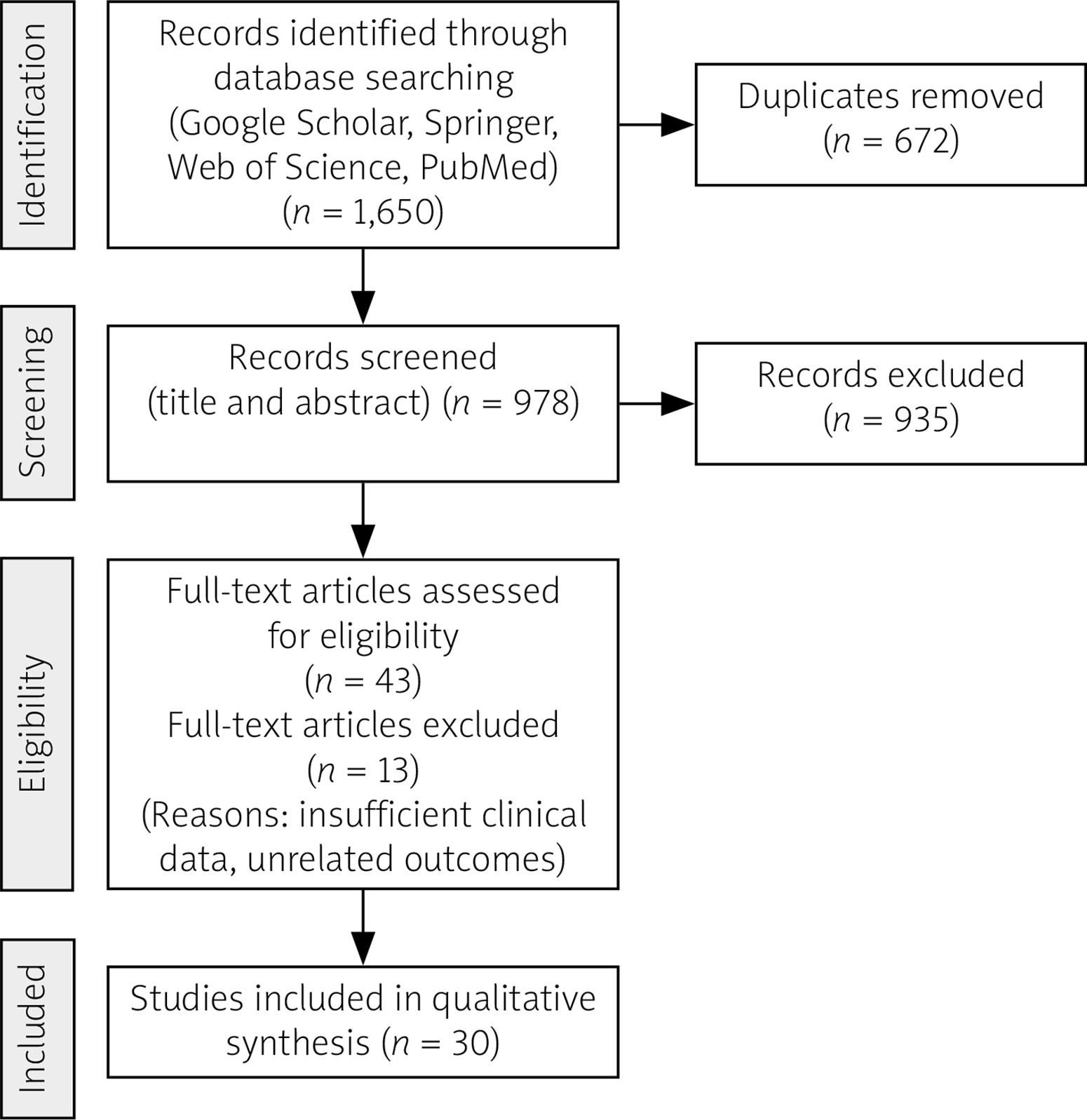
One thousand six hundred and fifty articles were identified in Google Scholar, Springer, Web of Science and PubMed. Only 30 research studies (one of them reported 8 cases) were included for review, as shown in Figure 1.
Case presentation
The case concerns a 19-year-old Saudi female patient with a known history of SLE, diagnosed 3 weeks prior at a private hospital based on inflammatory arthritis and pleural effusion (serositis), with positive anti-nuclear antibody (ANA) and anti-double-stranded DNA (anti-dsDNA) antibodies. Three days after the diagnosis, she had been admitted to another hospital for 5 days due to pleuritic chest pain and joint pain, receiving antibiotic coverage with GCs before being discharged. The patient was stable and had no further complaints. One day after discharge, patient experienced recurrent symptoms of chest pain and shortness of breath, prompting her visit to our emergency department. The patient was admitted under internal medicine as a case of pneumonia and started on intravenous (i.v.) antibiotics with rheumatology consultation to investigate the underlying cause of her symptoms, whether related to infection or disease activity.
Patient history
Upon reviewing the patient’s history, patient reported having pleuritic chest pain associated with shortness of breath upon exertion and hair loss for the last 3 days. The patient denied any history of headache, seizures, depression, blurred vision, double vision, malar rash, photosensitivity, Raynaud’s phenomenon, dry eyes, dry mouth, abdominal pain, or bleeding from other sites, and other systemic reviews were unremarkable. There was no family history of underlying connective tissue disease.
Physical examination
Upon examination, the patient was conscious, alert, and oriented. Vital signs were stable (blood pressure: 120/68, heart rate: 82, oxygen saturation: 96% on room air, temperature: 36.9°C). Chest examination revealed decreased air entry on the left side with crackles. Cardiovascular examination showed normal heart sounds (S1, S2 + 0). The abdomen was soft and lax, and there was no edema in the lower limbs. Systemic examination was otherwise unremarkable.
Clinical examination
The laboratory findings revealed several significant abnormalities suggestive of an underlying autoimmune and inflammatory disorder. The patient presented with marked anemia, evidenced by low hemoglobin (Hb; 8.1 g/dl), reduced serum iron (9.22 μg/dl), low total iron-binding capacity (62.15 μg/dl), and decreased transferrin saturation (15%), consistent with anemia of chronic disease or autoimmune hemolytic anemia. This was further supported by a positive Coombs test. Inflammatory markers were elevated, with an erythrocyte sedimentation rate (ESR) of 120 mm/h and a mildly elevated rheumatoid factor of 30 IU/ml, indicating an active systemic inflammatory process. The immunological evaluation revealed hypergammaglobulinemia (immunoglobulin G: 3,010 mg/dl) with low complement C4 levels and normal C3, and a normal thyroid profile, also favoring autoimmune etiology. There was an additional notable observation of severe vitamin D deficiency at 11 ng/ml, which is commonly found in chronic inflammatory conditions. Renal parameters were relatively preserved, with a mild reduction in creatinine (39 μmol/l), still within the normal reference range. Hence, overall findings indicate systematic active autoimmunity.
Imaging studies revealed bilateral pleural effusion, more pronounced on the left side, moderate pericardial effusion (PCE), and evidence of compression atelectasis on CT chest. A transthoracic echocardiogram confirmed normal left ventricular systolic function (ejection fraction > 65%) with no tamponade. These findings pointed to SLE activity with possible concurrent infection.
Chest X-ray findings and contrast-enhanced computed tomography of the chest
A chest X-ray was performed to assess the extent of pulmonary and pericardial involvement. As illustrated in Figure 2, the chest X-ray showed bilateral pleural effusion, with more fluid present in the left lung. Also, the patient underwent contrast-enhanced CT of the chest, which showed that the main pulmonary trunk and bilateral main branches appeared patent with no clear filling defect. The trachea and bilateral main airways were patent. No pneumothorax could be noted, with mild to moderate bilateral pleural effusion, more marked on the left side.
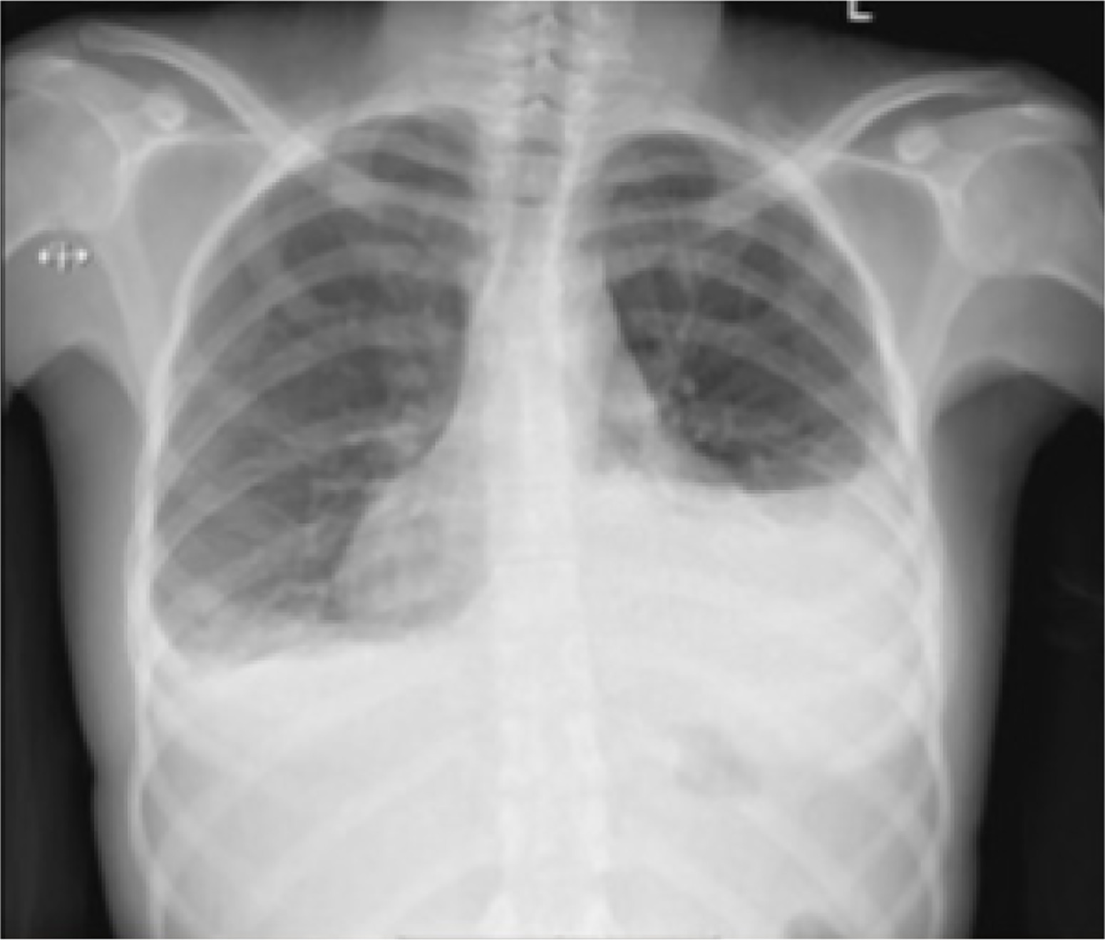
To sum up, the CT chest and X-ray findings were consistent with SLE-related serositis, presenting as moderate PCE and bilateral pleural effusions, more marked on the left. The associated basal lung atelectasis was likely secondary to compressive effects of the effusion. No evidence of pulmonary embolism, airway obstruction, or parenchymal consolidation was noted. These findings support a diagnosis of active SLE with pleuropericardial involvement and no current radiological evidence of infection or other complications.
Final diagnosis and impression
The rheumatology team’s impression pointed towards SLE activity, specifically serositis, possibly triggered by an underlying infection. Antibiotic coverage was initiated. Another differential diagnosis to consider is lupus pneumonitis, potentially secondary to infection or SLE activity. To determine the cause, the patient will be observed on antibiotics for 72 hours. If there is no response or deterioration, SLE activity is considered the more likely cause. Additionally, infection from Pneumocystis jirovecii pneumonia should be considered, particularly if the patient has been on high doses of GCs, although this information is not currently confirmed.
Treatment plan
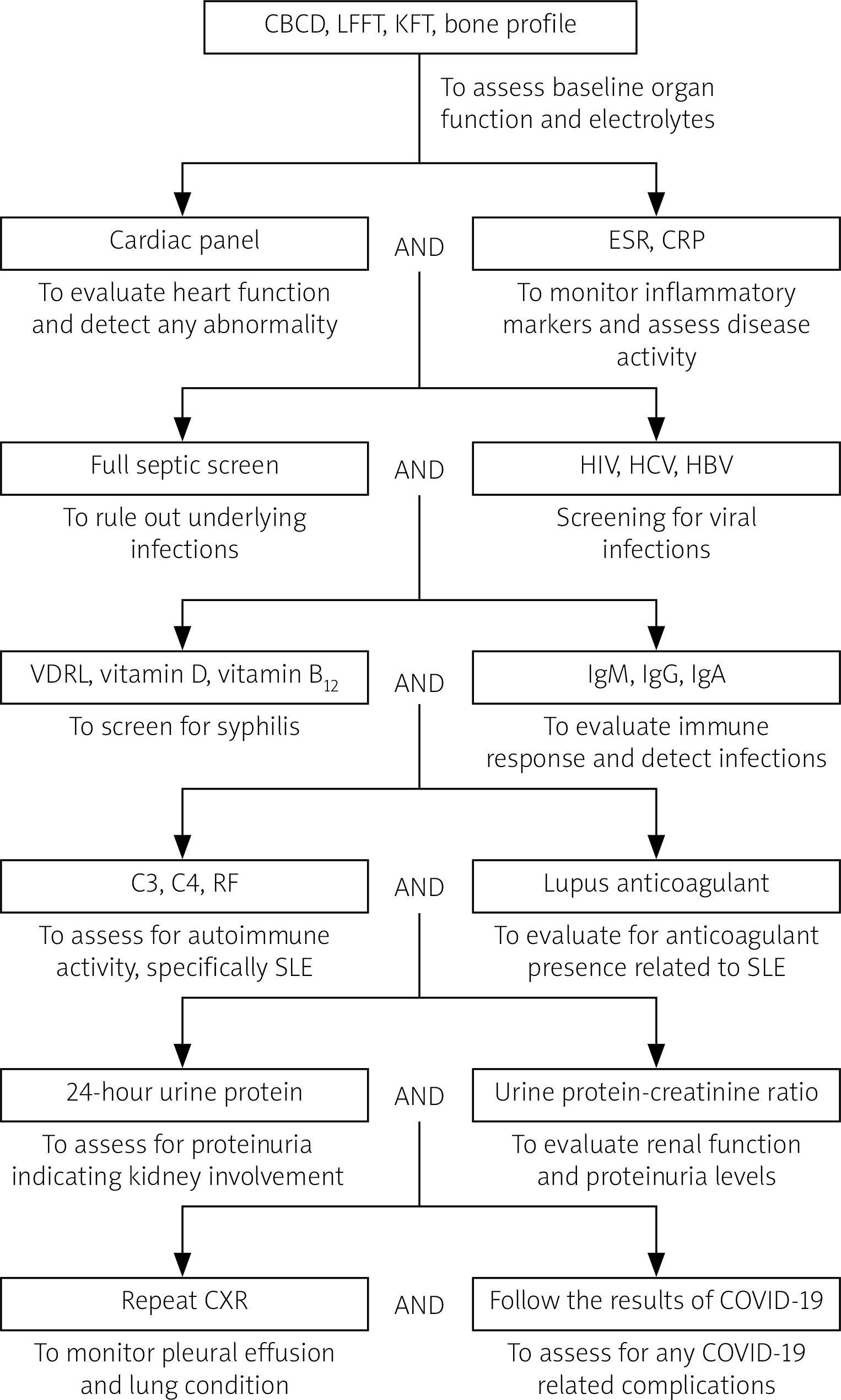
Figure 3 outlines a comprehensive treatment plan involving various investigations and procedures aimed at assessing organ function, detecting infections, monitoring disease activity, and evaluating immune and renal functions.
Fig. 3
Investigative algorithm for systemic lupus erythematosus-related pleuropericardial effusion.
C3 – complement component 3, C4 – complement component 4, CBCD – complete blood count with differential, COVID-19 – coronavirus disease 2019, CRP – C-reactive protein, CXR – chest X-ray, ESR – erythrocyte sedimentation rate, HBV – hepatitis B virus, HCV – hepatitis C virus, HIV – human immunodeficiency virus, IgA – immunoglobulin A, IgG – immunoglobulin G, IgM – immunoglobulin M, KFT – kidney function tests, LFFT – liver function tests, RF – rheumatoid factor, VDRL – Venereal Disease Research Laboratory test.
Pharmacological management
Patient was treated with methylprednisolone (500 mg i.v. for 3 days), hydroxychloroquine (HCQ; 200 mg p.o. twice daily), and supportive measures including calcium supplementation and intravenous immunoglobulins (IVIG; 1 mg/kg for 2 days), as shown in Figure 4.
Fig. 4
Therapeutic algorithm for pharmacological management of systemic lupus erythematosus (SLE)-related pleuropericardial effusion.
GCs – glucocorticosteroids.
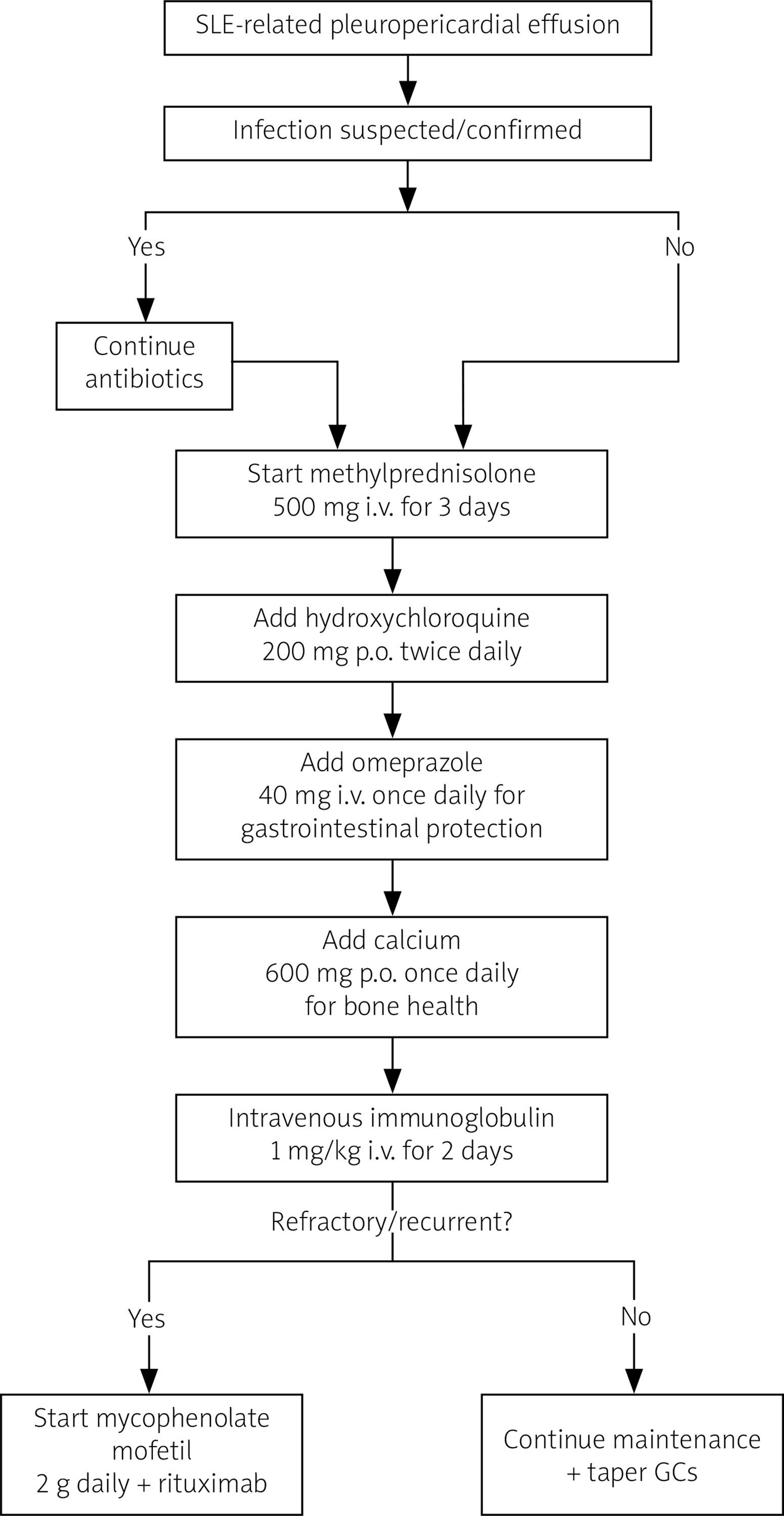
After starting the previously mentioned management, her condition improved significantly during the hospital stay, and she was discharged on a high-dose GC taper.
One month later, the patient presented with recurrent symptoms despite treatment adherence. In the rheumatology outpatient clinic, patient was started on MMF 2 g daily, with RTX considered as a potential escalation in therapy for refractory disease.
Discussion
This case illustrates a rare presentation of pleuropericardial effusion as the initial sign of SLE and highlights the difficulty in managing SLE-related serositis in a young patient. A 19-year-old female patient came to the hospital with bilateral pleural effusion, moderate PCE, and moderate activity of SLE according to laboratory markers including ESR, positive ANA, and anti-dsDNA antibodies. Even after treatment with GCs and antibiotics for presumed infection, her recurrence of symptoms highlighted the importance of differentiating between the infection and SLE activity in the patient. Immunosuppressive therapy is also highlighted in the management of the disease in ensuring disease control when she improved clinically following a high dose of GCs, HCQ, and IVIG, with the addition of MMF for refractory symptoms.
A study carried out in 2024 revealed an uncommon manifestation of SLE with cardiac tamponade and pleural effusion as apparent signs at the initial assessment. Their patient presented with severe dyspnea and systemic inflammation with both ESR levels and anti-dsDNA antibodies upregulated. Hence, GCs were used. This correlates with the pleuropericardial involvement in our case, but in terms of severity, our patient had no tamponade but had only moderate pleural effusion. However, both cases underscore the diagnostic challenge of distinguishing between SLE serositis and infection, a recurring theme in SLE management. The researcher focus on echocardiography for monitoring tamponade is reflected in our case, emphasizing its utility in evaluating effusion dynamics [6, 7].
In 2015, a group of scientists investigated pleural effusions associated with SLE and distinguished lupus pleuritis from non-lupus causes. They observed increased pleural fluid ANA titers (≥ 1 : 160) and strikingly low pleural fluid-to-serum complement ratios in lupus pleuritis, consistent with inflammatory activity. The unexplained pleural fluid ANA and complement tests were instrumental in ruling out non-lupus effusions and created a diagnostic path for the patient. While this case turned out positive for the Coombs test and low C4, the pleural fluid analysis that was performed in the study could have provided the much-needed diagnostic clarity. Glucocorticosteroids were effective, the scientists reported, with remission on these drugs mirroring that of our patient after GC treatment [8].
A study investigated the management of systemic inflammatory conditions such as adult-onset Still’s disease, emphasizing the role of interleukin (IL)-1 and IL-6 inhibitors alongside GCs. While the focus was not directly on SLE, the principles of managing systemic inflammation overlap with our case. The study advocated minimizing GC dependence to reduce adverse effects, an approach echoed in our patient’s transition to MMF for long-term management. The recurrent pleuropericardial effusion in our patient reflects the persistent inflammatory mechanisms reported by Cheo and Low [9], reinforcing the importance of targeted immunosuppressive therapy in chronic autoinflammatory conditions.
A group of scientists analyzed 5,679 pediatric SLE admissions, identifying 705 cases with PCE and noting higher mortality (2.4%) and readmission risks. This aligns with our case, where PCE marked severe SLE activity, but our 19-year-old patient responded well to GCs and HCQ without complications. This is in concordance with our case wherein PCE indicated severe SLE activity although the 19-year-old patient had a favorable response to GC and HCQ with no complications. Also, our patient had systemic inflammation with anemia (Hb 8.1 g/dl) and low C4, but she had a less severe disease duration with moderate pleuropericardial effusion and stabilization after early treatment. The pediatric population researcher had higher severity and the recurrence of symptoms. In contrast, our patient remained stable without requiring intensive intervention [10].
A 40-year-old female patient was diagnosed with SLE after having moderate PCE and exertional dyspnea. Diagnostic strategies include echocardiography, ANA, and positive anti-dsDNA as well as ruling out other causes including viral infections and malignancies. Methylprednisolone in high dose brought about complete improvement in symptomatology and disappearance of effusion. This case is in concordance with our case, primarily associated with pleuropericardial involvement and GC treatment which improved acute manifestations. However, due to the presence of effusion and disease activity in our patient, the clinical course was more severe and refractory, requiring treatment with MMF. In both cases, imaging and serological markers are discussed as tools for differentiating SLE activity from other causes of serositis [11].
Tocilizumab (TOCI), an IL-6 receptor antagonist, was used for managing recurrent, nonresponsive effusions involving the pleura and pericardium of a 22-year-old patient with SLE. The patient, whose condition did not improve with GCs and immunosuppressants, had no effusion after receiving TOCI, exhibiting the effectiveness of cytokine-targeted therapy in SLE-associated serositis. In contrast, our patient initially responded positively to GCs and HCQ, but recurrent symptoms required the introduction of MMF. The potential future treatment approach is biologic therapies in cases of refractory or recurrent serositis, providing a future pathway. Both cases highlight inflammation of serositis in SLE and the role of new immunosuppressive approaches [12].
In one report, a 47-year-old Iranian woman with massive PCE as the initial manifestation of SLE was diagnosed through ANA positivity, elevated anti-dsDNA, and Systemic Lupus International Collaborating Clinics (SLICC) criteria, and successfully treated with prednisolone, HCQ, and MMF over 6 months. While our patient presented with moderate pericardial and pleural effusion without tamponade, this case aligns with our case in highlighting the importance of immunosuppressive therapy in managing SLE-associated serositis. Our patient’s earlier need for MMF suggests a more refractory disease course. These cases underscore the role of echocardiography in monitoring effusion resolution and disease activity, despite differences in severity. Both cases emphasize the challenge of distinguishing lupus activity from infection [13].
In another case, an 11-year-old girl with pediatric lupus, who developed myocarditis, pulmonary hypertension, and a huge PCE, was treated successfully with methylprednisolone, IVIG, and CYC, entering long-term remission. Although our case was not complicated by myocarditis or pulmonary hypertension, it shares the inflammatory nature of SLE-associated serositis and the requirement for immunosuppressive therapies. Unlike the pediatric case that needed more advanced therapies, our patient did respond to GCs and HCQ, but with the recurrence of pleuropericardial effusion, advanced treatment options may need to be explored. Both cases highlight the difficulty of discriminating the activity of lupus from infection, which was addressed in our case by close monitoring and coverage with antibiotics [14].
In another report, a 35-year-old Sudanese woman, afflicted by a massive unilateral pleural effusion caused by SLE, as confirmed by systemic markers of inflammation and serological tests, required RTX after conventional treatment failed. Compared to this, our case showed bilateral pleural and moderate PCE with a good initial response to GCs and HCQ, implying a mild course. However, the recurrence of pleuropericardial effusion may be an indication for more aggressive biologics such as RTX, as used in the other case. Both cases highlight pleural effusion as a serious lupus manifestation with indications for aggressive immunosuppression and thorough diagnostic workup. The report stresses that in refractory or recurrent SLE-associated serositis, treatment escalation is crucial [15].
A pediatric case of pericarditis and PCE as the initial manifestation of SLE was described in a 7-year-old girl diagnosed through serological markers and echocardiography. Likewise, in our case, a 19-year-old patient with recurrent pleuropericardial effusion and SLE markers presented in a milder clinical state without severe systemic manifestations. Both cases were treated with GCs and appropriately tailored immunosuppressive therapy, using azathioprine in the pediatric case and MMF for our patient in view of the recurrence. The studies stress timely intervention, clinical imaging, and serological investigation, lessening age-related distinctions in the severity of the illness. Pediatric cases tend to need aggressive early immunosuppression due to the more severe initial presentation [16].
A 63-year-old woman with SLE was diagnosed via cytology of pleural effusion, experiencing chest pain, dyspnea, and recurrent effusions which resolved with GCs and HCQ. In our case, however, pleuropericardial effusion was ANA and anti-dsDNA positive, and SLE was confirmed on serological markers and imaging. Both cases highlight the inflammatory basis of SLE-associated serositis and the effectiveness of GCs, though our patient’s chronic symptoms necessitated MMF. These findings emphasize detailed diagnostics for atypical SLE and underscore the need for vigilant follow-up and advanced treatments for recurrent disease [17].
Another rare initial manifestation of SLE could be fibrinous constrictive pericarditis with large PCE, as has been recorded in a 36-year-old man who was found to have raised ANA and anti-dsDNA levels, confirming the diagnosis. The patient received treatment with non-steroidal anti-inflammatory drugs, GCs, and HCQ, and was free of symptoms in 6 weeks. Our patient had pleuropericardial involvement just as in that case; however, the effusion was of moderate quantity, without any fibrinous constrictive feature, and it proceeded to have a relapsing course after a brief remission with immunosuppressive therapy, thus requiring MMF therapy. These cases illustrate the importance of imaging, such as Doppler echocardiography, in diagnosing SLE-related pericarditis and effusion but also highlight the need to carefully rule out infection. The patient previously discussed improved rapidly; however, our case serves as a reminder that these patients may develop a recurrence requiring longer-term immunosuppressive treatment [18].
Severe cardiac compromise in new-onset SLE, including tamponade and antiphospholipid syndrome (APS), represents a grave autoimmune serositis manifestation. An 18-year-old girl with such a presentation needed emergency pericardiocentesis, GCs, and plasmapheresis in view of complications of respiratory failure. Both cases highlight the autoimmune nature of serositis and the importance of early diagnosis to avert severe outcomes, unlike our patient, who never experienced tamponade. Though our patient stabilized initially with GC therapy and HCQ, the later exacerbation of symptoms reflects the difficulties in treating refractory SLE. Complement monitoring (low C3 and C4) proved instrumental in evaluating disease activity in both instances. This emphasizes SLE’s aggregate systemic inflammatory burden, its overlap with APS, and the need for individualized multimodal therapy [19].
A patient with juvenile-onset SLE and treatment-resistant pleural effusion was, after only a modest response to GCs and MMF, successfully treated with TOCI, illustrating the importance of IL-6 in lupus pleuritis. Our patient, having bilateral pleural and PCE, responded first to GCs and HCQ and then later to MMF, showing that the illness course was less refractory. These 2 cases emphasize the inflammatory nature of SLE-associated serositis and support effective immunosuppression. In contrast to our case, managed according to standard protocols, the second report suggests IL-6 blockade as a potential option for persistent effusions. These cases, therefore, highlight the need for early intervention and later escalation to target therapy in refractory scenarios [20].
A 20-year-old woman with lupus myocarditis and PCE initially presented with left ventricular failure and was diagnosed with SLE upon ANA and anti-dsDNA antibodies. Since myocardial involvement is rare in lupus patients compared with pericarditis, early diagnosis and treatment with high-dose GCs significantly improved the situation. Our patient, on the other hand, developed moderate PCE with no myocarditis and cardiac failure but shared the diagnostic challenge of deciding between lupus activity and infection. The diagnosis in both cases was aided by echocardiography and biomarkers, with low complement levels indicating active lupus. The above-mentioned patient then responded well to GCs, whereas ours required MMF for recurring symptoms. This clearly delineates the distinct type of responses to treatment, which means that long-term monitoring and physician-guided treatment are warranted [21].
The initial SLE manifestation was myopericarditis and PCE in a 35-year-old female patient with complaints of chest pain, dyspnea, and systemic inflammation confirmed by elevated cardiac biomarkers, low complement levels, and positive ANA. High-dose intravenous methylprednisolone treatment led to symptom resolution, with no recurrence being reported following therapy. In contrast, our patient had SLE-associated serositis without myocardial involvement or elevated cardiac enzymes, subsequently being diagnosed with transthoracic echocardiography and laboratory markers for SLE-associated serositis and requiring further immunosuppressive therapy. Glucocorticosteroids as treatment and imaging for cardiac involvement were significant therapeutic considerations in both cases, suggesting that more advanced imaging methods such as cardiac magnetic resonance imaging may be useful for investigating future suspected myocardial involvement. Differences in clinical evolution and treatment outcomes were also observed in these cases [22].
The upcoming therapies are biologics, and they serve well in refractory SLE-associated serositis. A 51-year-old woman with SLE-associated pleuropericarditis not responding to standard treatment was able to achieve an extended remission period on belimumab, a B-cell-targeted biologic. This case highlights the role of biologics in managing refractory SLE-associated serositis, contrasting with our patient, who responded initially to GCs and HCQ but required MMF for symptom recurrence. Both cases emphasize early intervention and treatment escalation in refractory disease, with belimumab as a potential option if conventional therapies fail. The study underscores the evolving role of biologics in reducing disease activity and GC dependency, stressing personalized strategies for managing SLE-associated serositis [23].
Table II summarizes the characteristics of the presented cases (including the current case) and additional reported cases of SLE-associated pleuropericardial effusion, including clinical differences and treatment approaches.
Table II
Clinical characteristics, differences, and treatment outcomes of SLE-associated pleuropericardial effusion cases (including the present case)
| Author | Age, sex | Country | Initial presentation | Effusion type and severity | Key labs | Imaging findings | Treatment | Outcome |
|---|---|---|---|---|---|---|---|---|
| Bakhsh – current case (2024) | 19 F | Saudi Arabia | Chest pain, SOB | Bilateral pleural + moderate pericardial; no tamponade | ↑ ESR, ANA+, ↑ anti-dsDNA, low C4 | CXR and CT: bilateral pleural effusion; echo: moderate pericardial effusion | GCs, HCQ, IVIG, MMF | Improved; stable |
| Kavandi et al. [13] | 47 F | Iran | Dyspnea | Pericardial; massive | ANA+, ↑ anti-dsDNA | Echo: large effusion | GCs, MMF, HCQ | Resolved |
| Khatieb et al. [15] | 35 F | Sudan | Unilateral effusion | Pleural; massive, refractory | ANA+, ↑ anti-dsDNA | CXR: unilateral PE | RTX | Resolved |
| Chen et al. [14] | 11 F | Taiwan | Myocarditis, PAH, massive PE | Pericardial; severe | ANA+, ↑ anti-dsDNA, low C3/C4 | Echo: massive pericardial effusion | GCs, IVIG, CYC | Remission |
| Weich et al. [24] | 8 cases | South Africa | Dyspnea, chest pain | Large pericardial effusion (some with tamponade) | ANA+, anti-dsDNA+, low C3/C4 | Echo: large effusion; some tamponade | GCs + drainage | No recurrence |
| Alghamdi et al. [25] | 24 F | – | Dyspnea, chest pain | Pleural + pericardial; tamponade | ↑ ESR, ANA+, ↑ anti-dsDNA | Echo: tamponade | GCs | Resolved |
| Ocampo et al. [12] | 22 F | Canada | Recurrent effusions | Pleural + pericardial; moderate | ANA+, ↑ anti-dsDNA | Echo and CXR: recurrent effusions | TOCI | Resolved |
| Shalomi and Ramesh [26] | Adult F | – | Cardiac tamponade | Pericardial; tamponade | ANA+, anti-dsDNA+ | Echo: tamponade | GCs + pericardiocentesis | Resolved |
| Amro et al. [6] | 24 F | Saudi Arabia | Severe pulmonary hypertension + chest pain | Large pericardial effusion with early tamponade | ANA+, anti-dsDNA+, low complements | Echo: large pericardial effusion | GCs, diuretics, immunosuppressives | Resolved |
| Barradas et al. [27] | 37 F | – | Chest pain, SOB | Pericardial effusion; initial manifestation | ANA+, anti-dsDNA+ | Echo: moderate effusion | GCs | Resolved |
| Kriegel et al. [28] | Adult F | – | Empyematous pleural effusion | Sterile empyematous pleural effusion (associated with SLE) | ANA+, anti-dsDNA+ | CXR: pleural effusion | GCs; management of infection suspicion | Resolved |
[i] ANA – anti-nuclear antibody, anti-dsDNA – anti-double-stranded DNA, C3 – complement component 3, C4 – complement component 4, CT – computed tomography, CYC – cyclophosphamide, CXR – chest X-ray, ESR – erythrocyte sedimentation rate, F – female, GCs – glucocorticosteroids, HCQ – hydroxychloroquine, IVIG – intravenous immunoglobulins, MMF – mycophenolate mofetil, PAH – pulmonary arterial hypertension, PE – pulmonary embolism, RTX – rituximab, SLE – systemic lupus erythematosus, SOB – shortness of breath, TOCI – tocilizumab, ↑ – increased.
Study limitations and future implications
The patient’s treatment response varies, which is a limitation of this study. It suggests that the differing effects of GC regimens require individualized therapeutic approaches. On the other hand, duality in diagnosis – abnormal echocardiography and increased biomarkers – may fail to convey the very essence of lupus cardiac angina, thus requiring further diagnostic criteria. The future management of SLE-associated serositis is likely to emphasize early diagnosis and specific individualized treatment plans. A biologic agent such as belimumab may become an appropriate alternative for treating refractory cases and thus reducing GC use. Cardiac MRI may further refine diagnosis, and targeted therapies along with immunosuppressive agents will be refined through ongoing research to provide better treatment outcomes that offer better solutions to face the challenges of SLE, and to prevent recurrence of pleuropericardial effusions.
Conclusions
Pulmonary manifestations, especially pleuritis with pleural effusion, raise significant diagnostic challenges in SLE. Early recognition and differentiation from infectious to autoimmune causes are crucial for treatment. Treatment mainly consists of GCs in an autoimmune scenario. If the symptoms persist or recur, immunosuppressants should be considered. Such differences in therapeutic responses suggest an individualized approach. Early aggressive treatment to reduce disease activity and prevent recurrence is paramount.