Introduction
Ankylosing spondylitis (AS) is a chronic, progressive inflammatory disorder primarily characterized by inflammatory low back pain, often accompanied by peripheral arthritis, enthesitis, uveitis, spinal deformity, and ankylosis. The prevalence of AS varies geographically, ranging from 0.1% to 1.8% in Europe and approximately 0.24% in China [1, 2]. While genetic factors are widely recognized as significant contributors to AS pathogenesis [3], the specific molecular mechanisms and genetic determinants underlying the disease remain incompletely understood. Notably, human leukocyte antigen B27 (HLA-B27) and other genetic loci have been implicated in disease susceptibility, with HLA-B27 accounting for familial aggregation of AS [4]. However, HLA-B27 is estimated to contribute to no more than 50% of the overall genetic risk for AS [5, 6].
Recent genome-wide association studies (GWAS) in Chinese populations have identified additional susceptibility loci, including 2p15, 5q14.3, and 12q12 [7]. The single nucleotide polymorphism (SNP) rs4552569 at 5q14.3 is located between the hyaluronan and proteoglycan link protein 1 and epidermal growth factor-like repeats and discoidin I-like domains 3 (EDIL3) genes. The hyaluronan and proteoglycan link protein 1 has been associated with spinal osteophyte formation and disc degeneration in Japanese women [8], while EDIL3 modulates bone formation through inhibition of the Wnt signaling pathway [9]. Another SNP, rs17095830 at 12q12, resides within an intron of anoctamin-6 (ANO6), which encodes a multipass transmembrane protein with Ca²+-dependent phospholipid scramblase activity. The ANO6 has been implicated in osteoclast formation and bone remodeling [10, 11]. Recent studies have reported associations between ANO6 SNPs (rs4768085 and rs17095830) and both AS susceptibility and disease severity [12], although no such association was observed in a Taiwanese population [13].
To further elucidate the role of ANO6 in AS pathogenesis within the Chinese Han population, we conducted a large-scale case-control association study and investigated potential aberrations in ANO6 expression among AS patients.
Material and methods
Subjects
All participants provided written informed consent for the collection and utilization of their blood and tissue samples, and the study protocol was approved by the Ethics Committee of the Third Affiliated Hospital of Sun Yat-sen University (No. [2022]02-007-02). For preliminary screening of informative SNPs in the exon and transcription regulatory region of ANO6, 29 patients homozygous for the rs17095830G/G mutation and 30 healthy controls (HCs) were recruited. The validation cohort included 388 patients with AS who were heterozygous for the rs17095830A/G mutation and 511 ethnically matched controls. Additionally, 40 patients and 32 ethnically matched HCs were enrolled for reverse transcription-polymerase chain reaction (RT-PCR) analysis of messenger RNA (mRNA) in peripheral blood mononuclear cells (PBMCs). Immunohistochemical analysis of tendon tissue was conducted on samples obtained from 9 AS patients and 5 HCs during hip replacement or spinal orthopedic surgery. All AS patients met the 1984 revised New York criteria for AS and were diagnosed by at least 2 qualified rheumatologists [14]. Demographic data, including age and sex, as well as clinical manifestations such as family history of AS, age at disease onset, symptoms (e.g. dactylitis, peripheral arthritis), hip joint involvement, uveitis, enthesitis, and laboratory markers (e.g. erythrocyte sedimentation rate and C-reactive protein), were systematically collected.
Genotyping
Genomic DNA was extracted from peripheral blood samples using the standard salting-out method [15]. To identify candidate SNPs in the exonic and transcription regulatory regions of the ANO6 gene, PCR was performed with specific primers (Suppl. Tables I, II). The purified PCR products were subsequently sequenced using an ABI 3,700 automated sequencer. Single nucleotide polymorphisms were selected based on a minor allele frequency > 5% and a Hardy-Weinberg equilibrium (HWE) p-value > 0.01 in controls.
Ribonucleic acid preparation and reverse transcription-polymerase chain reaction
Total RNA was isolated from PBMCs using Trizol reagent (Invitrogen, 15596-026, Germany) following the manufacturer’s protocol. After DNase digestion, reverse transcription of total RNA was carried out using the PrimeScript RT Reagent Kit with gDNA Eraser (Takara, DRR047A, Japan). The polymerase chain reaction amplification was performed in a 20 μl reaction volume containing 40 ng of complementary DNA, 800 nM forward and reverse primers, and SYBR Premix Ex Taq II (Takara, DRR820A, Japan), in accordance with the manufacturer’s instructions. The PCR cycling conditions were optimized as follows: initial denaturation at 95°C for 30 seconds, followed by 40 cycles of denaturation at 95°C for 5 seconds, annealing at 60°C for 34 seconds, and extension at 72°C for 1 minute, with a final denaturation step at 95°C for 15 seconds. The following primers were used for amplification: ANO6 (F: 5´-CCTCAGTGTGATAGGCTTTGTCCA-3´, R: 5´-ACTGCAAAGACCAGGGTTCCA-3´) and β-actin (F: 5´-TGGCACCCAGCACAATGAA-3´, R: 5´-CTAAGTCATAGTCCGCCTAGAAGCA-3´). The specificity of PCR products was validated through melting curve analysis. Real-time PCR amplifications were conducted using a PRISM 7500 Real Time System (ABI), with all experiments performed in duplicate. The housekeeping gene β-actin was amplified to normalize the quantity of sample RNA. Relative gene expression quantification was determined using the ΔΔCt method (where Ct represents the threshold cycle), as previously described [16]. Additionally, PCR products were subjected to agarose gel electrophoresis for further analysis.
Immunohistochemistry
For immunohistochemistry, tendon tissues were fixed in 10% neutral buffered formalin for 12 hours at room temperature, subsequently embedded in paraffin, and sectioned following standard protocols. Tissue sections were deparaffinized and rehydrated using established procedures. To enhance immunostaining intensity, sections were heated at 95°C for 10 minutes in citrate buffer (0.01 M, pH 6.0). Sections were then incubated with the primary antibody overnight at 4°C. Following incubation, sections were washed 3 times for 3 minutes each in phosphate-buffered saline and processed with the ChemMate EnVision HRP (DAKO, China) according to the manufacturer’s instructions. Immunoreactive signals were visualized using the 3,3´-diaminobenzidine substrate, which imparts a brown color to the target protein. Tissue architecture was delineated by counterstaining with hematoxylin. Immunostaining scoring was performed by 2 experienced pathologists blinded to the subject group, with the final score calculated using the histoscore method [17]. Histoscore was calculated as the product of staining intensity (0–3: negative, weak, moderate, strong) and the percentage of positive cells (0–100%).
Statistical analysis
The genotyping accuracy of each SNP was assessed through HWE analysis. Genotype and allele frequencies in both AS patients and control groups were evaluated using the χ2 test to verify HWE. Allelic associations and haplotype analyses between cases and controls were performed using PLINK, with adjustments for sex, age, and geographic origin. The strength of associations was quantified as odds ratios (OR) per additional minor allele, accompanied by 95% confidence intervals. Linkage disequilibrium (LD) coefficients (D´ and r²) were calculated using Haploview 4.1 [18]. Relative gene expression quantification and histoscore data were presented as mean ± standard error, where n denotes the number of independent experiments. Statistical significance was determined using Student’s unpaired two-tailed t-test or analysis of variance (ANOVA), with p < 0.05 considered statistically significant, as analyzed using SPSS 17.0.
Results
Demographic data
In the initial screening cohort, which included 29 AS patients and 30 HCs, the mean age of AS patients was 28.0 ±7.4 years, with a male-to-female ratio of 4.8 : 1, while the control group had a mean age of 27.3 ±7.1 years and a male-to-female ratio of 6.5 : 1. In the validation cohort, the mean age of AS patients was 28.1 ±8.7 years, with a male-to-female ratio of 5.7 : 1, and the control group had a mean age of 28.4 ±5.5 years, with a male-to-female ratio of 4.2 : 1. No significant differences in age or sex distribution were observed between AS patients and controls in either cohort (p > 0.05).
In the RT-PCR analysis cohort, the mean age of AS patients was 27.00 ±5.57 years, with a male-to-female ratio of 15 : 1, while the control group had a mean age of 27.53 ±5.46 years, and a male-to-female ratio of 12.3 : 1. Again, no significant differences in age or sex distribution were detected between the groups (p > 0.05).
Identification of candidate single nucleotide polymorphisms
Sequencing analysis identified 9 SNPs within the transcription regulatory region of the ANO6 gene. Following stringent quality control measures, including exclusion of SNPs with HWE p-values > 0.01, minor allele frequencies < 5%, and missing data, 5 SNPs (rs79662606, rs75712006, rs76186361, rs17095830, rs80224086) were retained for further validation and analysis. A statistically significant difference (p < 0.05) in genotype frequencies was observed for these SNPs between the AS and control groups (Suppl. Table III).
Validation of single nucleotide polymorphisms in a larger population
The 5 SNPs were validated through PCR-based direct sequencing in a cohort comprising 388 AS cases and 511 HCs. Samples from 2 distinct stages were pooled to facilitate comprehensive analysis. All 5 SNPs were found to be in HWE. Genotype frequency analysis demonstrated statistically significant differences (p < 0.05) between AS cases and controls across the entire sample set. Notably, the frequency of all risk-associated loci was significantly elevated in AS patients compared to HCs (Table I).
Table I
Risk allele association analysis of the 5 single-nucleotide polymorphisms in anoctamin 6 in 417 ankylosing spondylitis cases and 541 controls
Identification of 5 haplotypes associated with ankylosing spondylitis in a Chinese Han population
Linkage disequilibrium analysis demonstrated strong pairwise associations among the 5 SNPs (Suppl. Fig. 1), prompting subsequent haplotype analysis. The analysis revealed significant associations between specific haplotypes and AS susceptibility. Notably, the haplotypes GTGCG (OR = 65.76, p = 2.684 × 1059), ATGTA (OR = 14.76, p = 1.168 × 10¹⁶), and ACGTA (OR = 5.02, p = 5.937 × 1026) exhibited higher frequencies in AS cases, suggesting their role as risk factors. Conversely, the haplotypes GTGTA (OR = 0.007, p = 4.625 × 10⁶) and ACATA (OR = 0.09, p = 4.081 × 1098) were more prevalent in controls, indicating potential protective effects (Table II). To further investigate the clinical implications, 2,144 AS patients with comprehensive clinical data were selected from our previous GWAS dataset [7]. These patients were stratified based on the presence or absence of the identified risk haplotypes. However, no significant association was observed between the risk haplotypes and AS phenotypic manifestation (Table III).
Table II
Significantly associated haplotypes for the 5 single-nucleotide polymorphisms. All haplotypes with a frequency < 0.03 were ignored in analysis. Loci analyzed: rs79662606, rs75712006, rs76186361, rs17095830, rs80224086
Table III
Analysis of the clinical manifestations and anoctamin 6 risk haplotypes in ankylosing spondylitis patients
Expression of ANO6 messenger RNA in peripheral blood mononuclear cells of ankylosing spondylitis patients and healthy controls
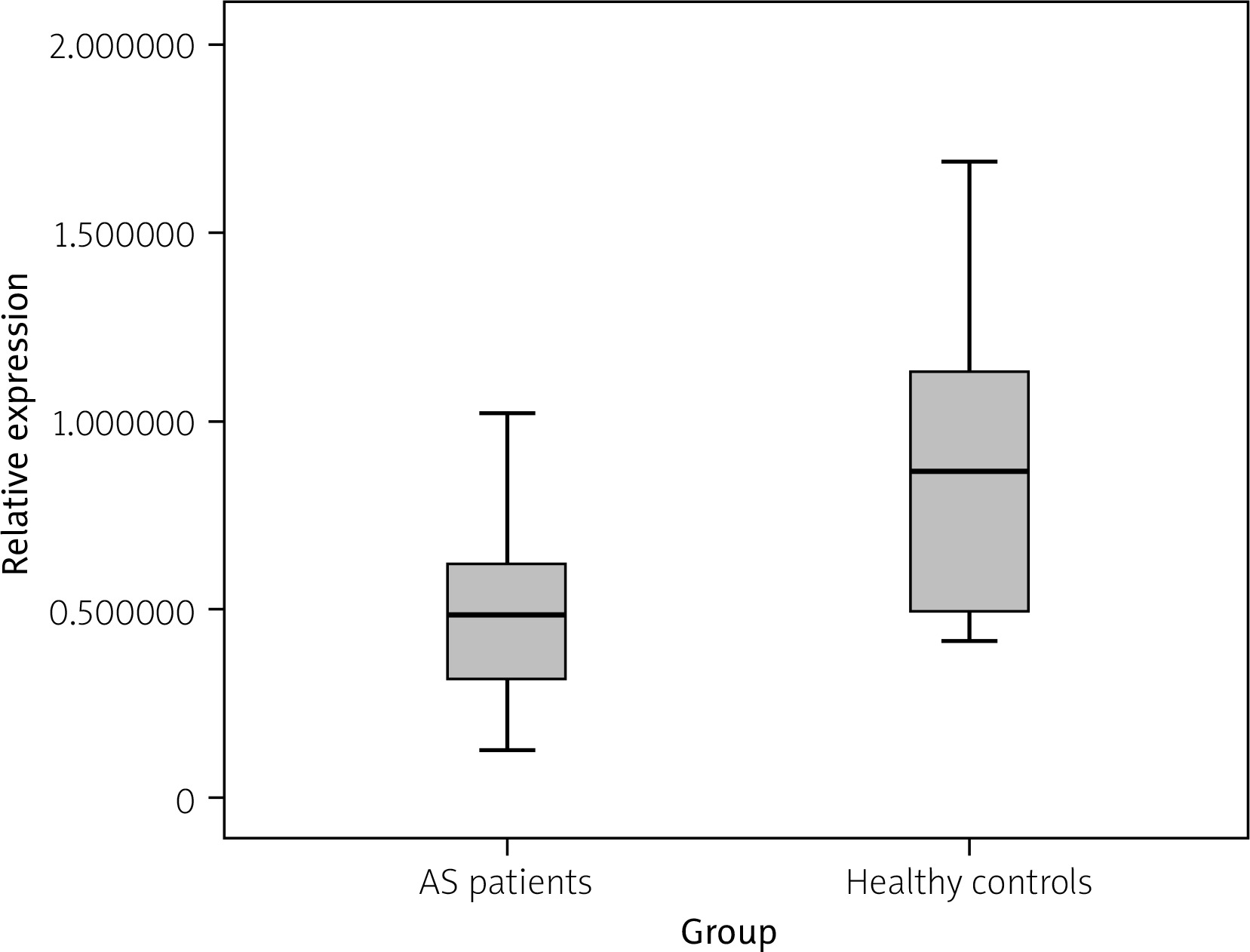
The expression levels of ANO6 mRNA were normalized to β-actin mRNA as an internal control. The mean relative expression of ANO6 in patients with AS (0.49 ±0.21) was significantly lower compared to HCs (0.86 ±0.38, p < 0.001; Fig. 1, Suppl. Table IV).
Expression of ANO6 protein in tendon tissue of ankylosing spondylitis patients and controls
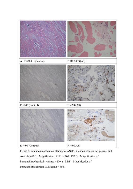
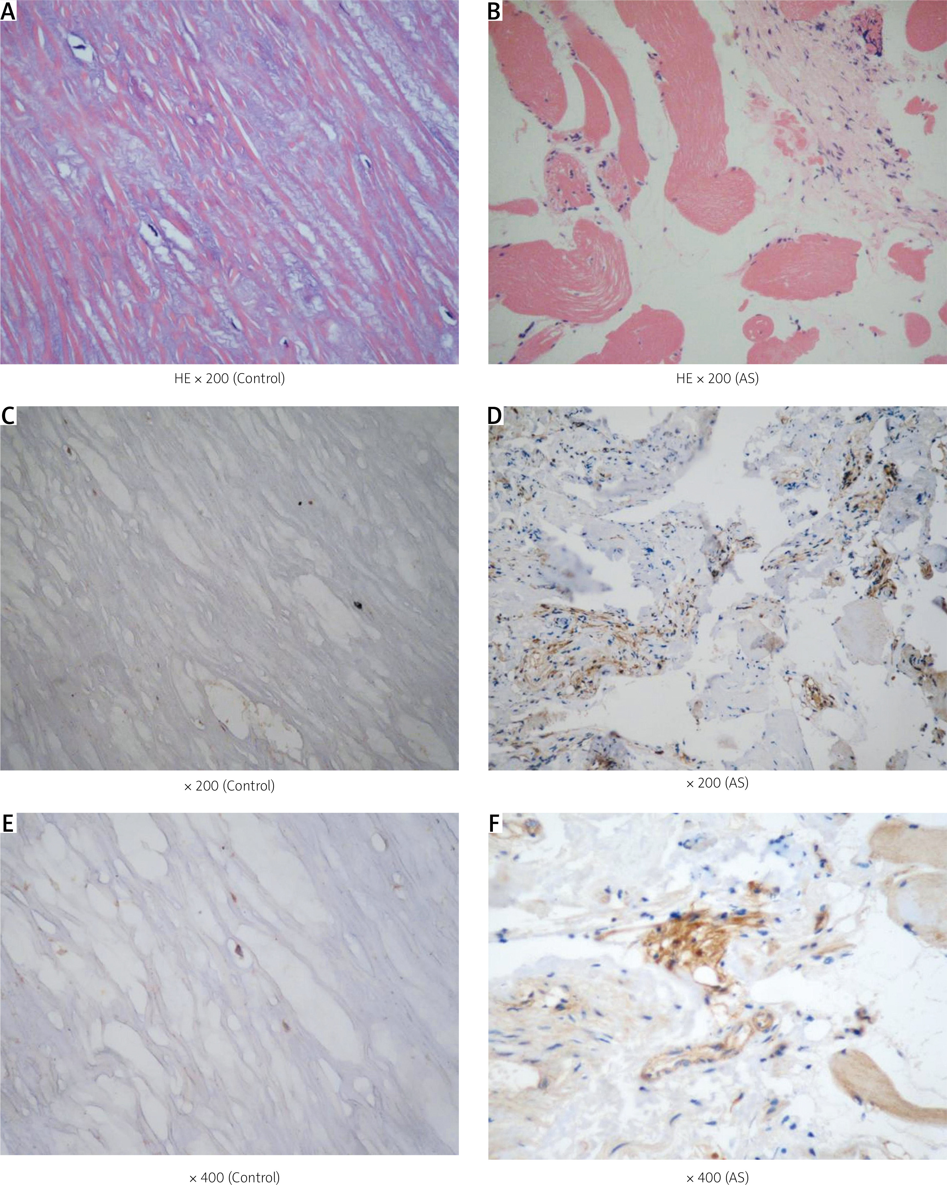
In ankylosing spondylitis patients, ANO6 protein was predominantly localized in myofibroblasts and lymphocytes (brown staining), whereas control tissues showed minimal or no staining (Fig. 2). In tendon tissue, the average histoscore of ANO6 protein expression, as determined by IHC, was significantly higher in 9 AS patients (182.22 ±30.732) than in 5 controls (62.00 ±35.637, p = 0.001; Suppl. Table V). The ANO6 protein was positively expressed in myofibroblasts, lymphocytes, and endothelial cells.
Fig. 2
Immunohistochemical staining of ANO6 in tendon tissue in ankylosing spondylitis (AS) patients and controls. A and B) Magnification of hematoxylin and eosin staining (HE): × 200. C and D) Magnification of immunohistochemical staining: × 200. E and F) Magnification of immunohistochemical staining × 400.
Discussion
Ankylosing spondylitis is widely recognized as a hereditary disease with polygenic involvement [19]. However, the mechanisms underlying aberrant bone formation and structural damage remain poorly understood, though they are likely influenced by multiple factors, including the localization of inflammatory lesions, entheseal stress, and the nature of the inflammatory response [20]. Following our initial GWAS identifying ANO6 as a susceptibility gene for AS [7], subsequent research reported that SNPs rs4768085 and rs17095830 in ANO6 were associated with AS susceptibility and disease severity [12]. However, no such association was observed for rs17095830 in a Taiwanese population [13]. To further investigate the genetic polymorphisms of ANO6 in relation to AS risk, we performed direct sequencing of exons and transcription regulatory regions in a Han Chinese cohort, followed by haplotype association analysis. Five SNPs with suggestive significance were validated in a larger Han Chinese population. All 5 SNPs within the transcription regulatory regions of ANO6 (rs79662606, rs75712006, rs76186361, rs17095830, rs80224086) demonstrated significant associations with AS, indicating a potential role of ANO6 in AS susceptibility in this population. Additionally, strong LD was observed among the SNP loci, and haplotype analysis revealed a higher prevalence of GTGCG (OR = 65.76, p = 2.684e-059), ATGTA (OR = 14.76, p = 1.168e-016), and ACGTA (OR = 5.02, p = 5.937e-026) in AS cases.
Single nucleotide polymorphisms in ANO6 may have direct functional implications in the molecular pathogenesis of AS. We hypothesize that the SNP located within the transcription control region could serve as a binding site for transcription factors, and that a specific combination of these factors may influence mRNA splicing modifications, ultimately affecting gene expression. Functional studies on ANO6 have demonstrated that interleukin-4 upregulates calcium-activated chloride channels during inflammation, which are regulated by ANO6 [21, 22]. In a murine model, ANO6 expression was observed in dendritic cells (DCs), and chemokine-induced migration of both immature and lipopolysaccharide (LPS)-matured DCs was attenuated following ANO6 knockdown [23]. Additionally, ANO6 activation is crucial for macrophage function [24]. Studies using knockout mouse models have revealed that ANO6 deletion results in a phenotype characterized by reduced skeletal size and deformities, evidenced by increased regions of non-mineralized, Ibsp-expressing osteoblasts in the periosteum during embryonic development and larger areas of uncalcified osteoid postnatally. Furthermore, primary ANO6(–/–) osteoblasts exhibit delayed mineralization, indicating a cell-autonomous role of ANO6 [25]. Notably, ANO6 expression is significantly elevated in mesenchymal stem cells compared to induced pluripotent stem cells or peripheral blood cells derived from axial spondylitis patients [26].
Ankylosing spondylitis is an inflammatory disorder associated with bone metabolism dysregulation. Emerging evidence suggests that ANO6, a protein implicated in phospholipid scrambling, may contribute to the pathogenesis of immunological processes and bone metabolism in AS, as supported by its association with disease susceptibility and severity in the Chinese Han population [12]. However, research on the functional role of ANO6 in AS and other autoimmune diseases remains limited. To address this gap, we conducted a preliminary study to compare ANO6 expression levels between AS patients and HCs at both mRNA and protein levels.
Reverse transcription-polymerase chain reaction analysis revealed a significantly lower relative mRNA expression level of ANO6 in PBMCs of AS patients compared to HCs. A hallmark pathological feature of AS is tendon inflammation. In 1972, John Ball first described the pathological characteristics of AS, noting that structural damage initiates at the tendon insertion sites, including ligaments, tendons, and joint capsules attached to bone, with particular emphasis on the significant involvement of the outer fibers of the intervertebral disc. Subsequent observations indicated bone loss followed by new bone formation, leading to osteophyte and syndesmophyte development [27]. Further studies identified infiltrations of T cells, B cells, bone marrow-derived macrophages, osteoclasts, and angiogenic cells within the tendon tissue insertion site, alongside the observation of inflammatory processes, bone destruction, bone formation, and other related phenomena [28, 29]. Semi-quantitative immunohistochemical analyses of tendon tissue revealed significantly elevated expression of ANO6, a gene encoding a calcium-activated phospholipid scramblase implicated in osteoclast differentiation and bone remodeling. Our findings suggest that risk haplotypes may disrupt ANO6-mediated phosphatidylserine exposure, potentially exacerbating inflammation-driven ectopic ossification in AS entheses [25, 26]. Given the multifactorial nature of AS and its impact on diverse physiological processes, it is plausible that ANO6 may contribute to AS pathogenesis through its involvement in tissue-specific mechanisms.
Study limitations
Among the study limitations, the limited sample size for tendon tissue analysis (9 AS vs. 5 controls) may have reduced the statistical power of the protein expression analysis. Larger cohorts are needed to confirm and extend these findings, including assessment of their generalizability to non-Chinese populations.
Conclusions
We identified disease-associated LD loci and haplotypes of ANO6 in AS and conducted a preliminary investigation into its expression. Our findings corroborate GWAS results, indicating that ANO6 gene polymorphisms are significantly associated with AS susceptibility in the Chinese population. These results provide novel insights into the complex interplay of genetic, immunological, and infectious factors in the etiology and pathogenesis of AS.