Introduction
Granulomatosis with polyangiitis (GPA) is an autoimmune small- and medium-sized vessel vasculitis. According to classification criteria, it belongs to the group of anti-neutrophil cytoplasmic antibody (ANCA)-associated vasculitides (AAV) [1]. It is characterized by necrotizing granulomatous inflammation and pauci-immune glomerulonephritis. The classical presentation typically involves a triad of organ systems: the upper respiratory tract (including the paranasal sinuses, nose, ears, and larynx), the lungs (pulmonary nodules, infiltrates, cavitary lesions, diffuse alveolar hemorrhage, bronchial stenosis) and the kidneys (rapidly progressive glomerulonephritis). However, GPA may impact nearly any organ system, complicating both diagnosis and management. Manifestations of central nervous system (CNS) involvement may include generalized symptoms such as headaches, meningitis, and seizures, as well as localized focal lesions such as stroke, cranial nerve paralysis, sensorineural hearing loss, and intracranial mass lesions [2]. Ocular and orbital manifestations are common in almost half of patients with either the limited or the systemic form of GPA [3]. Granulomatosis with polyangiitis can have a variable clinical course. It may have a localized form (often ANCA-negative) over extended periods of time, making recognition of the condition challenging for physicians lacking expertise in vasculitis. This type of AAV may remain localized or progress to a generalized form, causing damage to other organs. Infiltrative lesions may also spread locally, requiring extensive differential diagnosis. The diagnosis of GPA is based on clinical manifestations and complementary tests, including laboratory (detection of ANCA antibody), histopathology, and imaging studies. Histopathological confirmation is frequently unfeasible due to the complexity of acquiring tissue for microscopic examination and the presence of non-specific findings [4]. Although most patients with a clinical diagnosis of GPA are proteinase-3 ANCA positive, cases of myeloperoxidase (MPO) ANCA positivity and ANCA negativity have also been described. Although ANCA antibodies are involved in the pathogenesis of GPA [5], they may also be present in other conditions, e.g. connective tissue diseases, infectious diseases and drug-induced symptoms. Atypical perinuclear ANCA (pANCA) may be present in patients with inflammatory bowel disease and autoimmune liver diseases, including autoimmune hepatitis (AIH) [6]. They react primarily with MPO, less frequently with elastase, lysozyme, lactoferrin, cathepsin G, and catalase [7].
Here we describe the case of a female patient with AIH who developed a pseudotumor of the orbit followed by isolated cranial nerve palsies as a primary presentation of GPA. In addition, positivity for pANCA directed against elastase made it difficult to establish the correct diagnosis. The aim of this study is to raise awareness of this rare but significant clinical manifestation of GPA.
Material and methods
A literature search for patients with GPA manifesting as infiltration of the skull base presenting as multiple cranial nerves palsies was carried out using PubMed and ScienceDirect without date restriction. The search was conducted using the following key words in PubMed: (“Granulomatosis with Polyangiitis” [MeSH] OR “Granulomatosis with Polyangiitis”) AND (“Cranial Nerve Diseases” [MeSH] OR “Cranial Nerves” OR “Cranial Nerve Involvement”) AND (“Onset” OR “Initial Presentation” OR “Early Symptoms” OR “Isolated”). The following key words were used in ScienceDirect: (“Granulomatosis with polyangiitis”) AND (“Cranial nerve involvement” OR “Cranial nerve” OR “Cranial nerve disease”) AND (“Onset” OR “Initial presentation” OR “Early symptoms” OR “Isolated”).
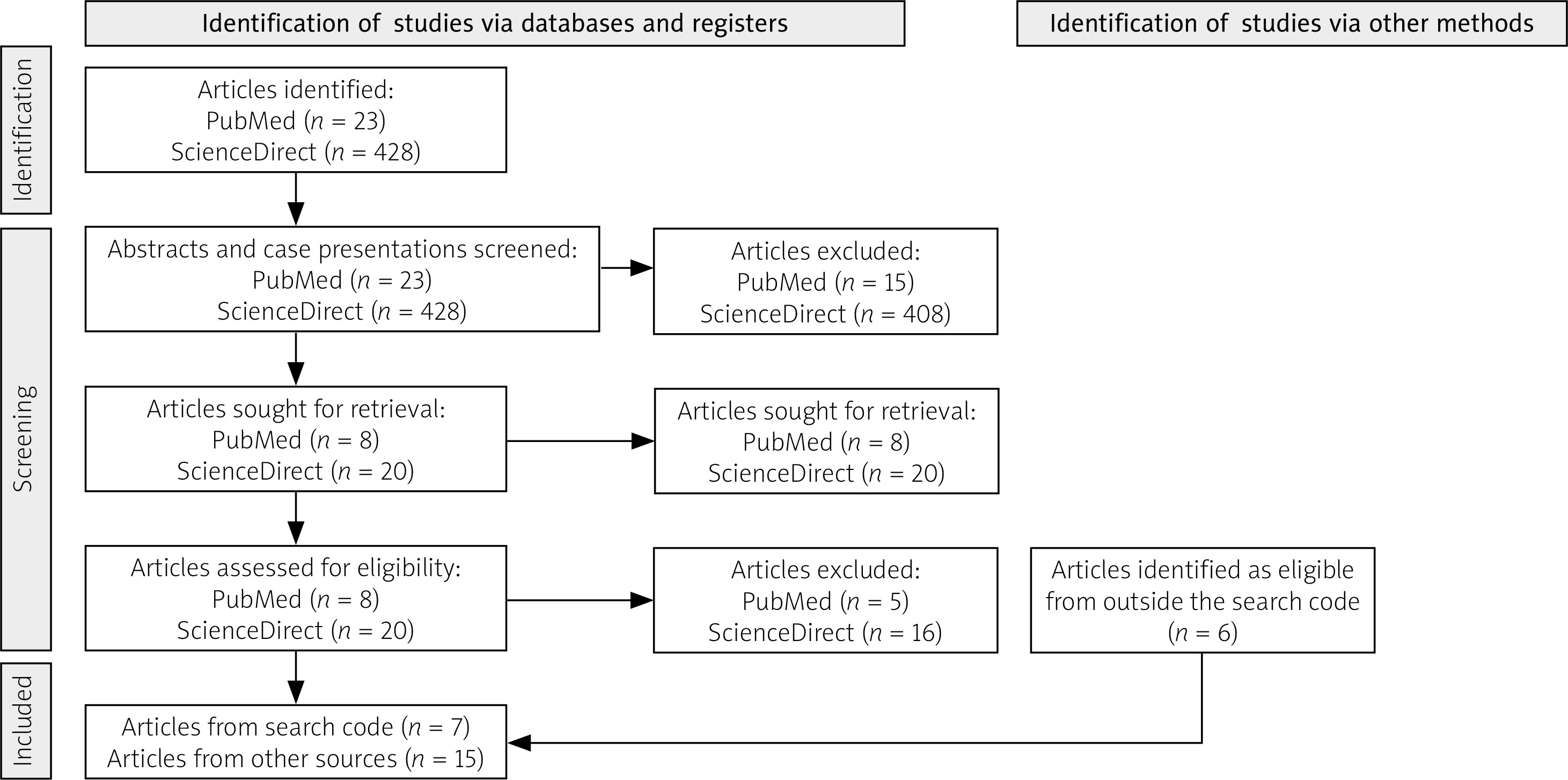
Using a combination of these search terms, we undertook a systematic review of the literature published in English, limited to full-text publications of original articles, letters to the editor, and case reports in peer-reviewed journals (the search strategy is presented in Figure 1).
This study was conducted in accordance with the principles of the Declaration of Helsinki from 1975 (revised in 2000), and written informed consent was obtained from the patient.
Search results
A review of the literature revealed only single reports of cranial nerve palsy as an initial symptom of GPA. Table I summarizes 4 case reports (aged 36–74 years; 3 men and 1 woman) with CNS involvement in the form of cranial nerve palsies. Otological and facial-orbital symptoms predominate: ear pain, hearing loss, VII nerve palsy, diplopia/limited eye movement, facial sensory disturbances, and taste and smell disorders. Most patients had multiple nerve involvement (including cranial nerves II, V, VI, VII, VIII, IX), and in 1 case the authors did not specify which nerves were affected. Imaging shows recurrent inflammatory/infiltrative changes in the sinuses and temporal bone (mastoid, tympanic cavity, thickening of the bone walls, lysis of the ossicles), sometimes with thickening of the dura mater or infiltration spreading to the cranial cavity; in 2 cases, changes in the lungs were also noted.
Table I
Review of cases with cranial nerve palsy as the initial manifestation of GPA
| Age, sex | Cranial nerve involvement | Initial symptom | Additional complaints | Findings on radiological imaging | Article |
|---|---|---|---|---|---|
| 74, female | Left-sided cranial nerve V3, VII, VIII, and IX palsies | Glossopharyngeal neuralgia | Left ear pain Bilateral hearing impairment Cloudy tympanic membranes Bilateral hypoesthesia of the skin below the mouth | Pseudotumoral nasopharyngeal lesion infiltrating cranial nerve V3 and intracranial invasion through foramen ovale Bilateral pulmonary infiltrates | Marini et al. [25] |
| 73, male | Cranial nerves not specified by the authors | Bilateral ptosis Redness of eyes Right facial weakness Right-sided hearing loss | Bilateral hyposmia Diminished right pupillary light reflex Limited right eye abduction Reduced sensation of the upper right facial region Dysgeusia Facial and bilateral orbital pain | Bilateral temporal thickening of dura Mucosal thickening of maxillary sinuses and bone wall thickening | Horiuchi et al. [26] |
| 63, male | Right-sided cranial nerve II, V, VI, VII, and VIII palsies | Right-sided facial weakness Diplopia Inability to close right eye Right-sided tongue ageusia | Right-sided hearing loss Right-sided blindness Paralysis of right abductor muscle Right maxillary sinusitis | Soft tissue densities in right mastoid area Osteoblastic activity in right mastoid bone and sphenoid bone Densities in lower lung fields | Daderian et al. [27] |
| 36, male | Left-sided cranial nerve VII palsy | Left-sided cranial nerve VII palsy Left-sided otalgia and hearing loss | Left-sided perforated eardrum with purulent effusion | Opacification of mastoid air cells and tympanic cavity with ossicular lysis | Batinović et al. [28] |
Case description
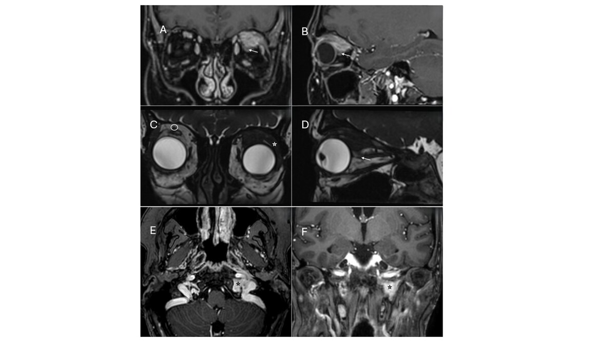
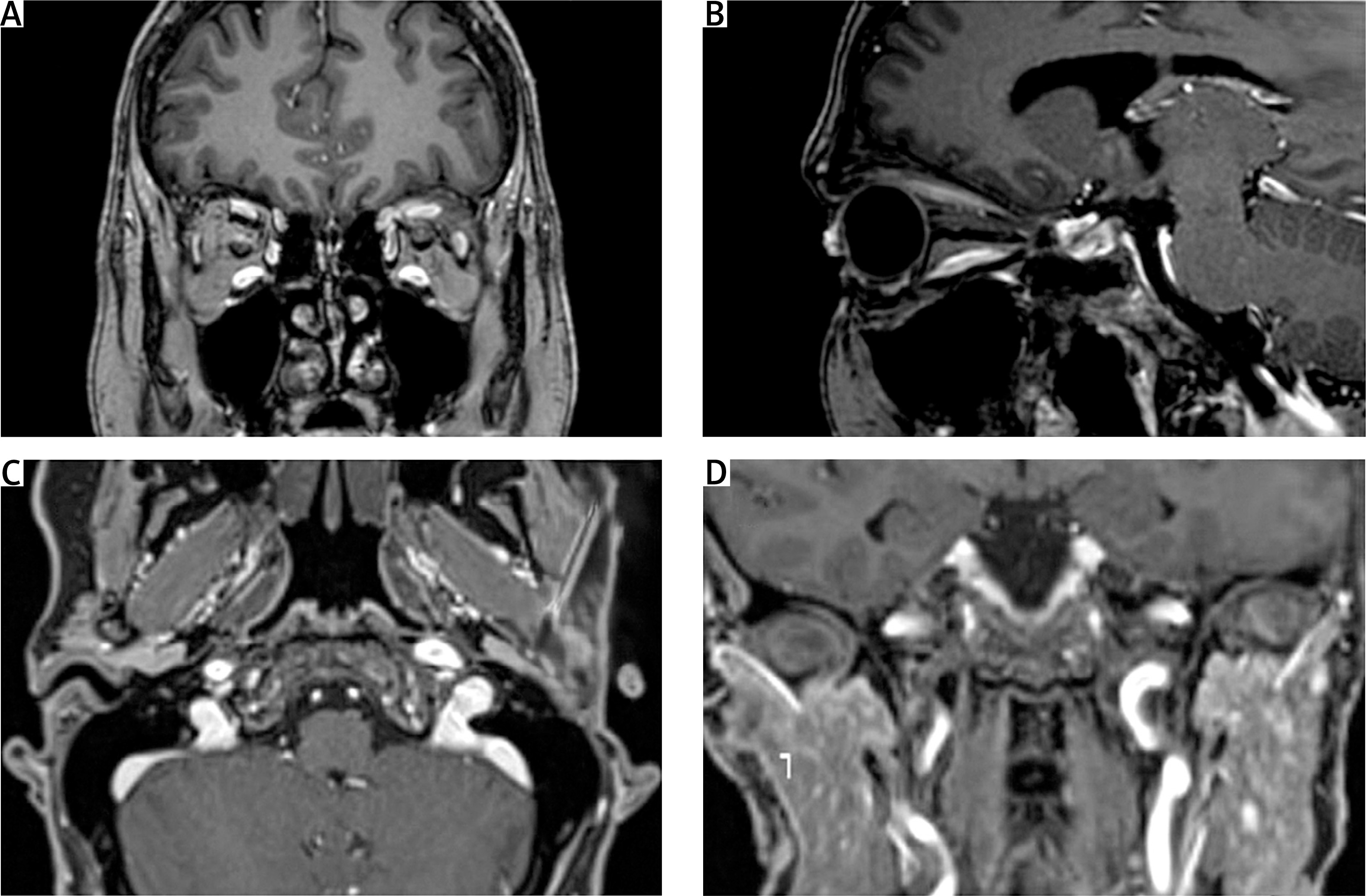
A 39-year-old female patient with cirrhosis of the liver due to AIH and autoimmune hypothyroidism was admitted to the rheumatology department for the presence of a pseudotumor of the left orbit with suspicion of an immunoglobulin G4-related disease (IgG4RD) for further diagnosis and management. Patient reported a 2-year history of ptosis of the left eye with left-sided numbness and flushing of the entire face. Magnetic resonance imaging (MRI) revealed a pathological mass in the upper part of the left orbit infiltrating the lacrimal gland, levator palpebrae superioris, superior rectus muscle and upper part of the lateral muscle. This mass extended deep into the orbit and was adjacent to the optic nerve just posterior to its exit from the globe (Fig. 2 A–D).
Fig. 2
Initial magnetic resonance imaging (MRI) showed a pseudotumorous, infiltrative lesion in the left orbit. The lesion enhanced after contrast administration (arrows in A, B) with infiltration of the lacrimal gland (asterisk in C), lateral rectus muscle, and superior rectus/levator palpebrae muscle complex, along with the supraorbital nerve. The lesion was in close proximity to the globe and the retrobulbar segment of the optic nerve (arrow in D), although these structures were not infiltrated. The supraorbital nerve is marked (circle in C) for reference. A brain MRI revealed a left-sided skull base infiltration at the level of the internal jugular vein foramen (asterisks in E, F) extending into the hypoglossal nerve canal.
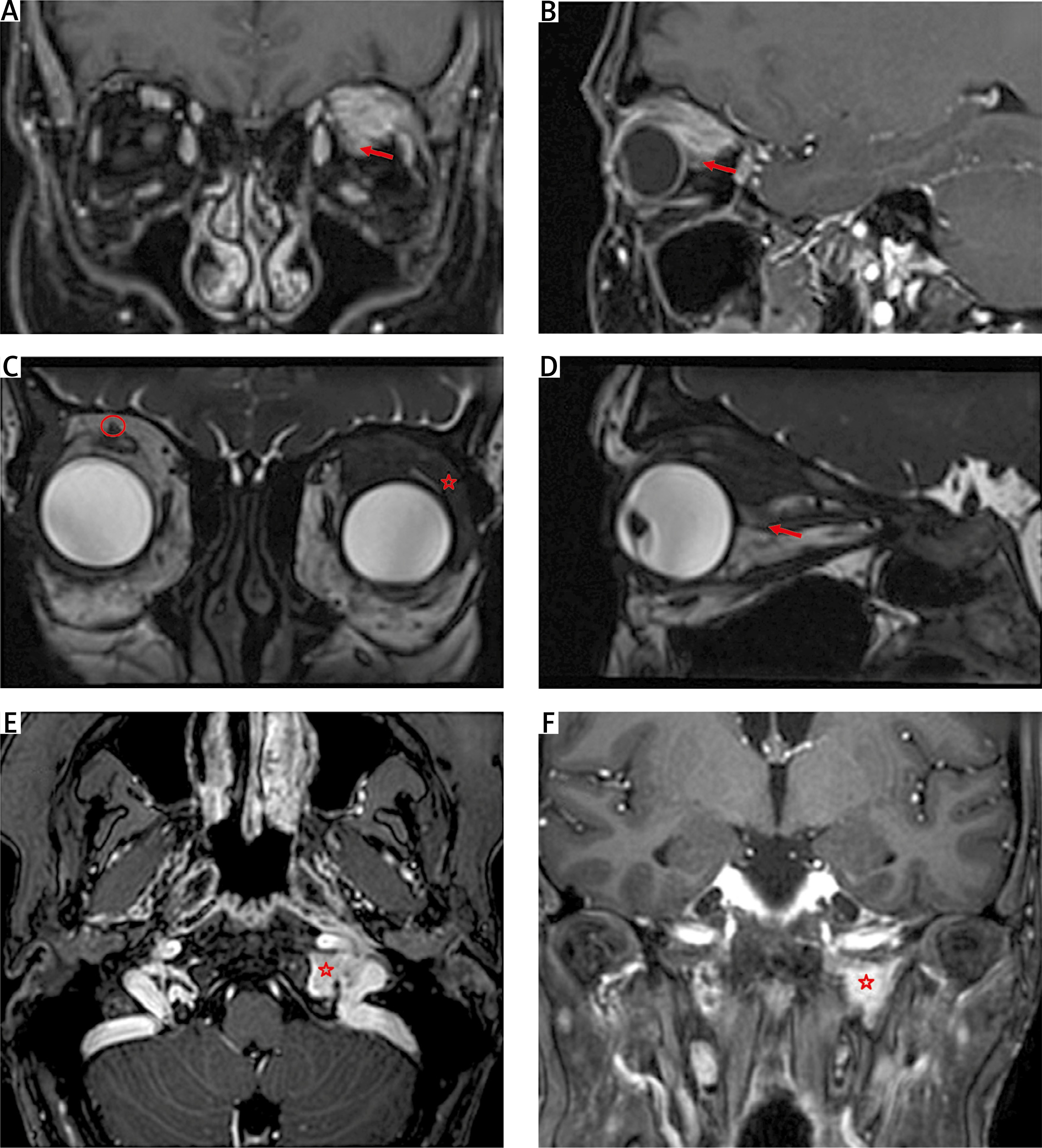
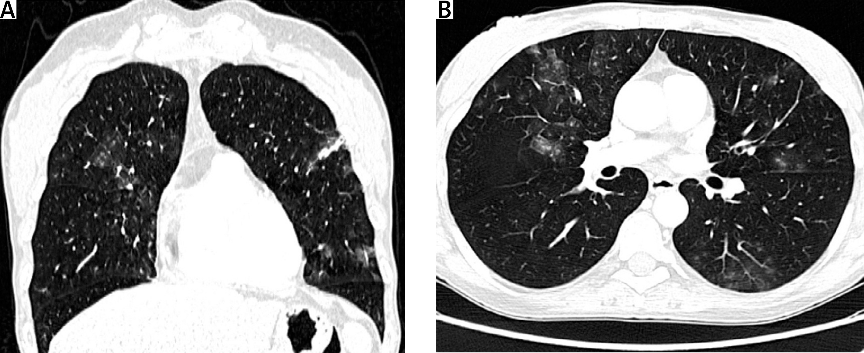
Systemic glucocorticosteroid (GC) therapy was initiated with prednisone 60 mg daily, which resulted in resolution of the symptoms. However, the symptoms returned when the steroid dose was tapered. A biopsy of the orbital mass revealed only non-specific collagenous fibrous tissue. The patient experienced gradually increasing weakness, decline in physical performance, significant weight loss, dysphagia and hoarseness. The patient denied hypersensitivity to ultraviolet radiation, diarrhea, constipation, dysuria, cough, shortness of breath, chest/abdominal pain, swelling, and runny nose. On physical examination, the patient had left eyelid ptosis with associated swelling of the periorbital tissues. The neurological examination revealed discrete symptoms of damage to the ophthalmic branch of the trigeminal nerve (V1) and cranial nerves IX–XII on the left side. The consulting laryngologist diagnosed inflammatory changes in the larynx, paralysis of the left vocal fold, paresis of the right vocal fold, absence of reflexes from the posterior pharyngeal wall, and large retention of secretions in the lower pharynx. Laboratory tests revealed microcytic anemia (hemoglobin 10.7 g/dl; mean corpuscular volume [MCV] 78 fl), slightly elevated inflammatory parameters (C-reactive protein [CRP] 16 mg/l, normal < 5 mg/l), and presence of antinuclear antibodies (antinuclear antibodies human epithelial type 2 > 1 : 2,560, homogeneous + fine-grained type). The immunoblot profile showed positive p/SSA Ro-52 antibodies. The presence of pANCA 1 : 2,560 pANCA was also found, corresponding to anti-elastase antibodies in the ANCA profile. The liver immunoblot profile was positive for Gp210 (anti-glycoprotein-210 antibodies), antibodies to soluble liver antigen/liver pancreas, anti-SSA Ro-52 antibodies, and anti-smooth muscle antibodies 1 : 320 (normal < 1 : 20). Despite the absence of pulmonary symptoms, chest computed tomography revealed, in the upper lobes of both lungs and in segments VI and X of the left lung, ground-glass opacities and a few areas of consolidation that could correspond to changes in the course of vasculitis (Fig. 3). Magnetic resonance imaging of the brain showed a left-sided skull base infiltration located between the bulb of the internal jugular vein extending into the hypoglossal nerve canal (Figs. 2 E, F). Due to the initial suspicion of IgG4RD, serum immunoglobulin G and immunoglobulin G4 (IgG4) levels were tested, but both were within the normal range. Additionally, IgG4 staining was performed on a liver biopsy obtained during the diagnosis of AIH showing staining in isolated plasmacytes, while a lacrimal gland biopsy exhibited negative staining. The neurosurgeon decided against diagnostic sampling of the posterior cranial fossa due to high procedural risk. Based on the comprehensive clinical evaluation along with immunological, radiological, and histopathological findings (negative IgG4 staining), a diagnosis of pANCA-positive vasculitis was established. Immunosuppressive treatment with methylprednisolone was administered (methylprednisolone 3 g i.v. in total), followed by prednisone therapy (1 mg/kg/day, 50 mg/day with a gradually reduced dose). Therapy with cyclophosphamide (CYC) at a dose of 15 mg/kg according to the European Vasculitis Study Group regimen was initiated. The patient’s clinical condition quickly improved after intense immunosuppressive therapy. She received 9 infusions of CYC with a cumulative dose of 8.42 g. Six months after initiating treatment, the patient showed remission of orbital and skull changes on MRI (Fig. 4), with a Birmingham Vasculitis Activity Score of 0. Cyclophosphamide was discontinued, and azathioprine (AZA) was initiated as maintenance therapy. Currently, 5 years after the start of treatment, the patient is in remission. Treatment continues with AZA 100 mg and low-dose prednisone (5 mg/day).
Discussion
Granulomatosis with polyangiitis is a multi-organ systemic autoimmune disease that manifests with a variety of symptoms. Clinical features may result from both inflammatory granuloma formation and small vessel inflammation. In addition to the classic presentation of upper respiratory, pulmonary, and renal symptoms, eye and nervous system involvement are among the symptoms associated with GPA. Ocular and orbital manifestations are present in almost half of patients with either the limited or the systemic form of GPA. Frequently, they may be present as the initial features of the disease [1]. The clinical spectrum of ophthalmic symptoms is highly variable, since almost every part of the eye and orbital structures may be affected. Inflammation of the conjunctiva, cornea, episclera, sclera, retina, lacrimal gland, and orbital blood vessels leads to a diverse range of symptoms, including ocular pain, erythema and edema of the eyelids, conjunctival injection, nasolacrimal duct obstruction, limited extraocular muscle movements, proptosis, diplopia, and vision loss [8]. The occurrence of an orbital mass is less frequent in patients with GPA. In most patients, such a mass manifests with reduced vision, diplopia, and proptosis. In a study that included 1,142 patients with GPA, only 5% developed orbital masses during a 5-year follow-up. An orbital tumor may be the sole location of the disease (limited form of GPA) or may coexist with involvement of other organs [9]. The differential diagnosis of orbital infiltration must consider a wide spectrum of diseases, including infections, neoplasms, and other inflammatory orbital diseases such as idiopathic sclerosing orbital inflammation, thyroid or Graves orbitopathy, sarcoidosis, as well as IgG4RD (Table II) [10–17]. The non-specific fibrous tissue found on histopathology of the orbital infiltrate biopsy in our patient initially suggested IgG4RD. However, the lack of elevated serum IgG4 levels and negative IgG4 staining on biopsy, as well as positive ANCA and lung and skull involvement, favored the diagnosis of GPA.
Table II
Differential diagnosis of orbital and skull base infiltration
[i] ACE – angiotensin-converting enzyme, CRP – C-reactive protein, CT – computed tomography, GCs – glucocorticosteroids, GPA – granulomatosis with polyangiitis, IgG4 - immunoglobulin G4, IGRA – interferon-γ release assay, MALT – mucosa-associated lymphoid tissue, MRI – magnetic resonance imaging, TB – tuberculosis, TSH – thyroid-stimulating hormone, TRAb – thyroid-stimulating hormone receptor antibodies.
Central nervous system involvement in GPA has been reported in 7–11% of cases [18]. Cranial nerve palsy occurs in 2–10% of cases. Wójcik et al. [19], based on the Polish vasculitis registry, stated that CNS involvement affects approximately 10% of patients with GPA. Similarly, Bonek et al. [20] estimated the frequency of cranial base involvement to be 3–10% of GPA, with a potentially severe disease course and resistance to treatment. Symptoms of CNS involvement may result from a variety of lesions: a granulomatous disease spreading from the ear, nose, and throat to neighboring structures, direct meningeal involvement due to adjacent granulomatous disease in the skull base, paranasal or orbital region, or the formation of a granuloma primarily in the nervous system [21]. In the present case, the disease began with an orbital pseudotumor, but it was not until the onset of cranial nerve palsy that the diagnosis was made and appropriate treatment initiated. Interestingly, the patient had no symptoms of lung involvement, and the lesions were visualized only by computed tomography. This indicates the need for a multi-organ assessment of every patient with GPA regardless of reported symptoms.
Skull base infiltration refers to the spread or invasion of a tumor, infection, or inflammatory condition into the skull base, the bony structure at the base of the skull. This can lead to various symptoms and complications depending on the underlying cause and the extent of the infiltration. Causes of this clinical state include neoplastic processes such as lymphoma, primary bone tumors, and metastatic lesions. However, these would not normally present with raised inflammatory parameters. Other causes include inflammatory pseudotumor, osteomyelitis, tuberculosis, sarcoidosis, as well as fibrous dysplasia and Paget disease. Table I provides detailed information on differential diagnosis of skull base and orbital infiltration. Kiessling et al. [2] conducted a retrospective analysis of 29 patients with skull base GPA. Out of the patients 72% presented skull base symptoms as the initial manifestation of the disease. They also concluded that 83% of the studied patients at some point experienced cranial neuropathies. De Groot et al. [22] analyzed the neurological manifestations of GPA and concluded that in most cases, the cranial nerve involvement is a result of infiltration of granuloma from the paranasal sinuses. In their study, 6 out of 128 patients had cranial nerve involvement. A cohort study conducted by Wojciechowska et al. [23] compared clinical signs of GPA and microscopic polyangiitis (MPA). Out of 41 patients with GPA, 8 presented with nervous system involvement, 3 of which had cranial nerve palsies. Interestingly, all cases with nervous system involvement were in the course of GPA. Tsuzuki et al. [24] analyzed the clinical profiles of GPA in 16 patients; 1 patient presented with hearing loss, tinnitus and paralysis of cranial nerves VII, IX, and XII, and another patient presented with otalgia and paralysis of nerve VII.
The following studies report cases in which cranial nerve dysfunction was the initial symptom and further investigation resulted in the diagnosis of GPA. Detailed information on each case is provided in Table II. Marini et al. [25], Horiuchi et al. [26], and Daderian et al. [27] all described patients who had a history of several cranial nerve palsies for a few weeks. The patients were ultimately diagnosed with GPA after radiological imaging, laboratory tests, and biopsy. Batinović et al. [28] described a patient who experienced acute otitis media complicated with facial nerve palsy. The diagnosis of GPA was confirmed after finding elevated cytoplasmic ANCA (cANCA) and histological findings of transbronchial biopsy.
Clinical and experimental data demonstrate that ANCAs play a crucial role in the pathogenesis of AAV [29]. Depending on their immunofluorescence pattern on ethanol-fixed neutrophils, ANCAs were termed cANCA, pANCA, and atypical ANCA [5]. Perinuclear ANCA predominantly targets MPO. However, in non-vasculitic autoimmune diseases, pANCAs are often directed against other antigens such as elastase, lactoferrin, and cathepsin G [30]. In a study by Roosendaal et al. [6], ANCAs were found in 74% of patients with AIH. Due to the well-documented occurrence of pANCA in association with autoimmune liver diseases, our patients’ autoimmune profile, characterized by AIH and pANCA targeting elastase, was initially consistent with the underlying AIH. However, the development of skull base infiltration, cranial nerve palsies, and pulmonary infiltration indicated progression to a systemic vasculitic process, ultimately leading to the diagnosis of GPA.
Skull base infiltration with neurological symptoms is a potentially organ/life-threatening manifestation of GPA requiring rapid and intensive immunosuppression. According to the European Alliance of Associations for Rheumatology guidelines, treatment with a combination of GCs and either rituximab or CYC is recommended for patients with new-onset or relapsing GPA with organ-threatening or life-threatening disease, in order to induce remission. Avacopan in combination with rituximab or CYC previously recommended for induction of remission in GPA, as part of a strategy to substantially reduce exposure to GCs was withdrawn by the FDA and EMA based on an analysis of the registration clinical trial data and reported adverse events, and the article from the clinical trial published in NEJM was retracted [31].
Conclusions
Cranial nerve palsy as an initial manifestation of GPA is most likely caused by cranial nerve compression by an inflammatory tumor of the skull base. Considering our case alongside the existing literature, we note that cranial nerve impairment is not a common manifestation of GPA. Given the small number of cases, a typical clinical presentation cannot be defined. However, this manifestation is important to recognize, since it may lead to delayed diagnosis and disease progression if overlooked. Our findings highlight that all patients with suspected GPA, regardless of the presented symptoms, require a comprehensive multi-organ assessment. Larger patient cohorts are needed to better characterize this rare presentation and facilitate earlier diagnosis in such cases.