
Introduction
Psoriasis (Ps) is a chronic, systemic inflammatory disease affecting 0.09–11.4% of the population according to the “Global report on psoriasis” presented in 2016 by the World Health Organization (WHO). The most common type (up to 80% of all cases) of this disease is plaque Ps; erythrodermic, pustular, flexural, guttate, and follicular types occur much less frequently [1, 2]. The etiology of the disease is not fully understood. Genetic, environmental, and immunological factors play a substantial role in its pathogenesis. Hypotheses suggest that the disease is triggered in genetically predisposed patients by environmental factors, such as bacterial, viral, and fungal infections, as well as alcohol, tobacco, stress, certain medications, and injuries [1, 3–9]. Genetic factors make the disease occur in a hereditary manner in about 30% of the cases. The mode of inheritance seems to be multifactorial and polygenic [10–13]. The genetic studies identified fifteen Ps-susceptibility regions, known as PSORS1–15 [6, 13]. Psoriasis has been found to be associated with human leukocyte antigen class I (HLA), including HLA-B13, HLA-B17, HLA-B57, and HLA-Cw6. Interestingly, HLA-Cw6 is associated with more severe skin changes, but less with psoriatic arthritis (PsA) development [6, 13–15]. Genetic studies have shown that the HLA-Cw2 and HLA-Cw6 alleles appear to increase PsA susceptibility, while HLA-Cw4 may have a protective effect [16]. The association of HLA-B27 antigen and uveitis is well documented, and its presence may predict development of arthritis, sacroiliitis, and uveitis in Ps patients [1, 13, 17].
Outline of immunological system dysfunctions in psoriasis
The basic pathological mechanism responsible for the development of Ps is a dysfunction in the Th lymphocyte system, which leads to uncontrolled secretion of inflammatory cytokines, including tumor necrosis factor (TNF) and interleukins (IL) – IL-17 and IL-22 [1, 3, 14, 15, 17–20]. However, dendritic cells also play a major role in the initial stages of the disease as antigen-presenting cells. Their activation and production of interferons (IFN) α and β, as well as the activation of type I IFN signaling, promote Th1 and Th17 differentiation and activity including IFN-γ and IL-17 production. Th17 cells produce IL-17, IL-21, and IL-22, which activate keratinocyte proliferation [15, 21–23].
The proliferation of keratinocytes is stimulated by the inflammatory environment, in particular the activity of TNF, IL-17, and IFN-γ. Keratinocytes themselves are involved in the inflammatory process through the secretion of pro-inflammatory cytokines (IL-1, IL-6, and TNF), chemokines, and antimicrobial peptides (AMPs), such as LL37, β-defensins, and S100 proteins [18]. The TNF–IL-23– Th17 pathway is particularly important in the etiopathogenesis of plaque Ps. In view of the above, currently available biological therapies are directed primarily at inhibiting described pathways (anti-TNF, IL-12/IL-23, IL-17). They also target the Janus kinase/signal transducers and activators of transcription (JAK/STAT) pathway, which are pleiotropic cascades interacting with other signaling pathways as a regulatory mechanism [19, 20, 24, 25].
Clinical picture of psoriasis
Psoriasis is a chronic, immune-mediated inflammatory disease that manifests itself in a variable manner, in various morphologies (hyperkeratotic, pustular, or mixed) and in various body localizations [7].
Skin Ps is generally divided into plaque Ps (the most common), guttate Ps (second most common), pustular, erythrodermic (severe form, unstable plaque Ps), and inverse Ps. Depending on the age of onset and clinical features, we also divide Ps into two basic types. Type I develops before age 40 and type II after age 40 [7].
The differences between the two main subtypes of Ps are presented in Table I [1, 17].
Table I
Psoriasis is often associated with several diseases such as PsA, obesity (abdominal or central), diabetes mellitus type 2, and cardiovascular disease. Quite often, symptoms of depression may occur [21, 26].
The prevalence of PsA in the psoriatic group is estimated at up to 30%. Psoriatic arthritis is a heterogeneous disease that may affect either the axial (spine and sacroiliac joints) or peripheral skeleton, and patients often show overlapping domains. Because of this variability, clinical presentation can range from isolated enthesitis or dactylitis to widespread peripheral arthritis or predominant axial involvement, making diagnosis and management challenging. Notably, skin changes usually precede joint involvement, but there is no strict association between the skin disease activity and arthritis [10, 11, 27, 28].
The GRAPPA group (Group for Research and Assessment of Psoriasis and Psoriatic Arthritis) recognizes five key clinical subtypes (domains) of PsA, reflecting the heterogeneous nature of the disease: 1) peripheral arthritis (both asymmetric oligoarthritis and symmetric polyarthritis); 2) axial disease (sacroiliitis and spondylitis); 3) enthesitis; 4) dactylitis; and 5) skin and nail involvement [27]. The GRAPPA domain-based classification directly guides treatment: therapy is chosen according to the dominant clinical domain. Thus, treatment in PsA is personalized and domain-specific rather than uniform across all patients. In the GRAPPA framework, uveitis is considered an important extra-articular domain that significantly influences treatment selection in PsA; the presence of uveitis shifts biologic preference toward monoclonal TNF inhibitors (TNFi), which have the strongest and most consistent evidence for controlling ocular inflammation. This recommendation is consistent with European Alliance of Associations for Rheumatology (EULAR) recommendations for the management of PsA with pharmacological therapies (2023) [27, 28].
In patients with Ps and/or PsA, ophthalmological symptoms may be a symptom of the underlying disease or a coincidence; therefore, an ophthalmological consultation should always be considered to deepen the diagnosis and exclude potential causes (including infectious ones).
Eye involvement in psoriasis
Eye involvement in Ps is usually discussed in the context of PsA or wider spondyloarthropathies (SpA) and co-occurring uveitis. However, more attention should also be paid to other ocular symptoms emerging in the course of Ps.
According to various authors, ocular symptoms occur in 10–67% of patients with PS and in 31% of patients with PsA. More than one eye complication is present in 20% of cases of Ps, with both eyes being affected in 48% of such cases [29–31]. All parts of the eye may become affected in the course of Ps [32, 33]. The most frequently detected ocular pathologies include eyelid involvement, blepharitis, keratoconjunctivitis sicca (dry eye disease [DED]), conjunctivitis, corneal lesions, uveitis, episcleritis, and cataract [3, 14, 17–19, 34–39]. Table II presents the risk factors of eye involvement in Ps.
Table II
Ocular symptoms appear most often during the disease exacerbation; for this reason, they can be observed first by a rheumatologist or dermatologist rather than an ophthalmologist [17, 23, 40].
There is a statistically significant correlation between the occurrence of ocular symptoms and the severity of the disease assessed by the Psoriasis Area Severity Index (PASI) score. Significantly more often these symptoms are observed among patients with PASI > 5, with an average PASI of 17 at the time of diagnosis [33, 34, 37]. Studies have also shown that ocular symptoms are more common among patients with nail involvement, pustular Ps, PsA (particularly polyarticular type), as well as among patients treated with methotrexate (MTX) or acitretin [3, 14, 19, 41, 42]. However, in most studies no correlation between the occurrence of ocular complications and duration of the disease, age, or sex was found.
However, it should be emphasized that recurrent inflammation of eye structures causes gradual tissue damage, which leads to scars, visual disturbances, and even loss of vision.
It should also be noted that one of the most recent studies has documented the protective effects of biological treatment against corneal changes and Meibomian gland dysfunction in Ps [33].
All patients with Ps and PsA should undergo ophthalmologic consultation annually. Patients with active PsA and active skin Ps with PASI of 10 or above should undergo ophthalmologic consultation biannually.
Eyelid involvement
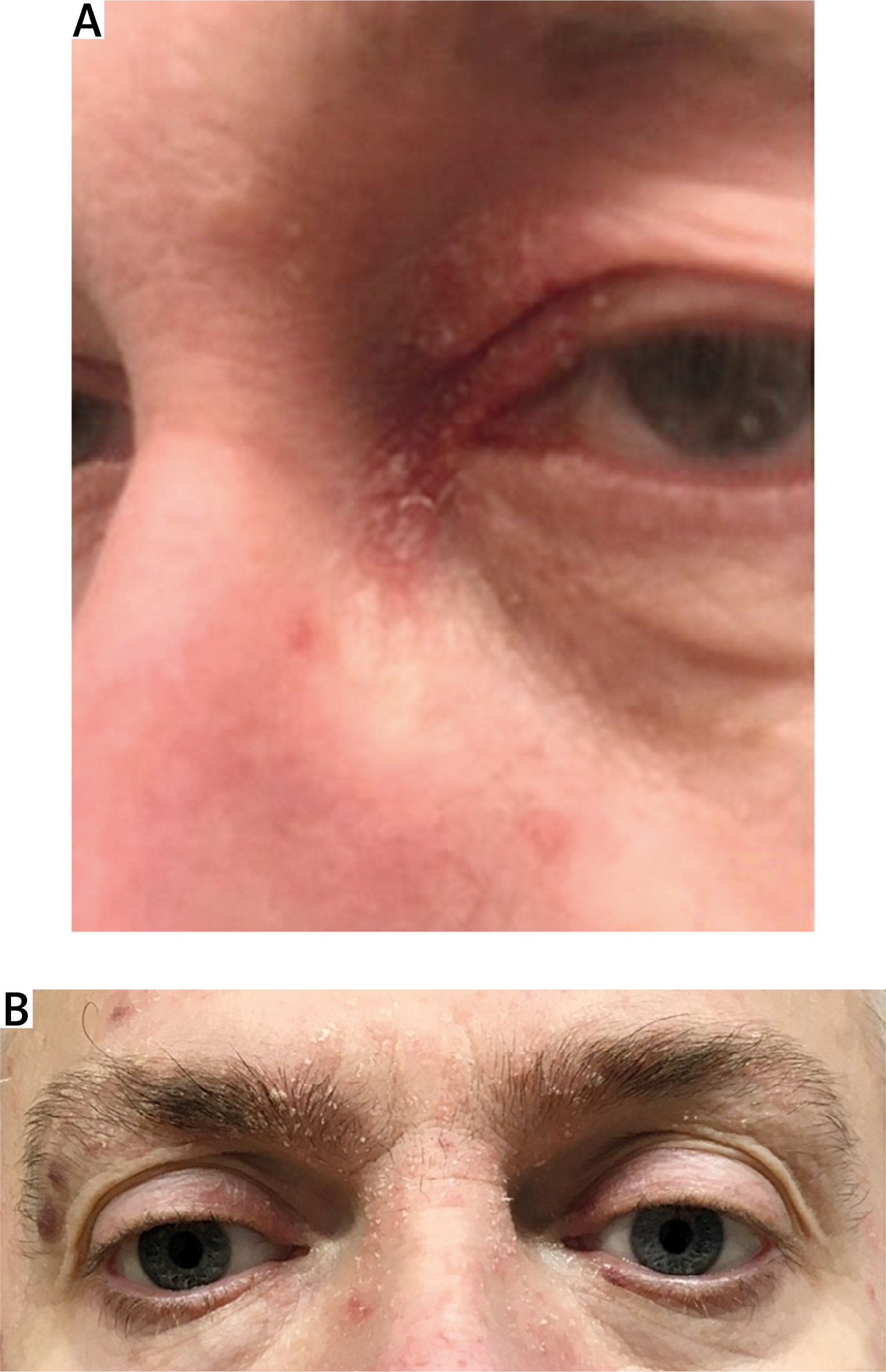
The face and particularly the eyelids are a rare localization of Ps. Facial manifestations of Ps with involvement of the eyelids, nasolabial folds, and perioral area are relatively rare; in these seborrheic areas, sebo-Ps can be expected to emerge more often than Ps vulgaris (Fig. 1) [43]. Such a manifestation is more common among patients with severe disease, protracted disease, and those with a family history of Ps. Topical vitamin D analogs and calcineurin inhibitors (tacrolimus, pimecrolimus) can be used in the treatment [12, 44, 45].
Fig. 1
Eye and facial manifestation of Ps: A) psoriatic changes in the angle of the eye; B) seborrheic form of facial psoriasis with changes on the eyelids and with blepharitis.
Recently, there has been growing evidence that these medications are also effective and safe for the treatment of eyelid inflammation and even result in improvement among patients with corneal changes in the course of Ps [12, 44, 46].
Blepharitis
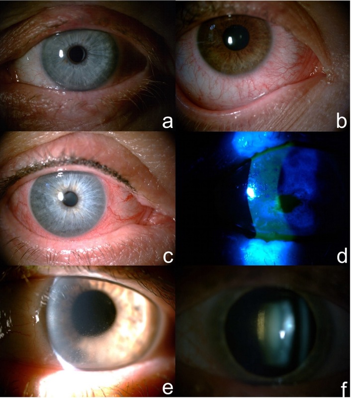
Blepharitis is the most common ocular complication of Ps (Fig. 1). It is caused by hyperkeratosis, Meibomian gland dysfunction, and mechanical obstruction of the Meibomian glands or lacrimal duct [3, 32, 34, 37]. Overgrowth of Staphylococcus aureus is also involved in the pathogenesis of blepharitis.
The patient may report symptoms of red eye, the sensation of having a foreign body in the ophthalmic region, severe eye burning, or intolerance of contact lenses [23, 37]. During physical examination it is worth paying attention to dandruff occurring mainly at the base of the eyelashes and red, swollen eyelids [1, 34]. The eyelid margins may also be affected by ulceration and crust formation.
The most important aspect in the treatment of blepharitis is proper hygiene, which consists of using warm compresses, as well as mechanical washing of the eyelids using specially designed cleaners or baby shampoo [1, 34, 37, 44]. In addition, a weak topical glucocorticosteroid (GC) may be used for a short period. Topical antibiotics are particularly useful for treatment of acute inflammation [37, 38].
Untreated eyelid inflammation may lead to contact lens intolerance, trichiasis, madarosis, cicatricial ectropion, and even visual impairment [1, 37, 39].
Dry eye disease
According to the latest research, DED affects 20 to 37% of patients with Ps and is significantly more common in Ps than among the healthy population [14, 17, 19, 39].
This syndrome is most likely the result of a disorder in the production of the tear aqueous component and tear film instability, as confirmed in studies using Schirmer’s test (without anesthesia and with anesthesia) and the tear break-up time (TBUT) test. Symptoms of DED include dryness, foreign body sensation, burning increasing during daytime, red eyes, and flakes on eyelids [29, 31, 34].
Artificial tears (drops or gel) are commonly used for the treatment (preferably medications without preservatives). It is also necessary to use ophthalmic ointments with paraffin before sleep in order to relieve nocturnal symptoms [1, 34]. Immunosuppressive therapy (cyclosporine drops) or eye drops containing the patient’s serum are used in severe cases of eye dryness. Lacrimal punctum occlusion is used in those cases with low lacrimal secretion (Schirmer test less than 5 mm/5 minutes). Relatively new methods of treatment include meibomian gland stimulation and electrostimulation of tear secretion [35].
Conjunctivitis
The incidence of conjunctivitis among patients with Ps is 10–13%, which is well above corresponding figures for the general population [3, 17, 19, 23]. A large percentage of patients with Ps presents in a conjunctival cytology examination a squamous metaplasia at mild or moderate stage [33]. Conjunctivitis can coexist with or can be caused by DED and eyelid inflammation [36, 47].
It manifests with pain, foreign body sensation under the eyelids, tearing, burning, and conjunctival injection [29–31, 37]. Mucopurulent discharge occurs in a bacterial infection, whereas bilateral watery discharge together with swollen preauricular lymph nodes indicates viral etiology. For a presumed bacterial etiology, artificial tears and antibiotics are recommended.
Corneal involvement
Corneal problems are found in 4–16% of cases, more often among patients with PsA and are usually secondary to DED, conjunctivitis, or blepharitis. This indicates the importance of treatment of these symptoms for the prevention of corneal involvement [29, 41]. Histological changes such as parakeratosis, inflammatory cell infiltrates, and angiogenesis are similar to psoriatic lesions on the skin or in joints, which suggests the involvement of common primary mechanisms [1, 44]. The most common corneal pathologies include punctuate epithelial keratitis, opacities, erosions, neovascularization, and scarring. Significant pain, photophobia, and blurred vision indicate corneal pathology, which requires urgent ophthalmologic consultation. In the Lee et al. [41] study, there was an increased risk of keratopathy in Ps patients even without preexisting prominent corneal disease. The authors concluded that this risk increases with exposure to Ps [41].
Uveitis
Uveitis is a serious complication occurring among 0.7–2.6% of patients with Ps and 7–9% of patients with PsA [43, 44, 48]. Studies have confirmed elevated levels of TNF, IL-2, IL-6, and IL-17 not only in patients with Ps but also in the aqueous humor of patients with uveitis, suggesting common pathogenetic pathways for these diseases [49].
In patients with Ps, uveitis most often affects the anterior part of the eye (anterior uveitis) and is characterized by a more chronic course, recurrence, tendency to occur bilaterally, and a more frequent need for non-steroidal anti-inflammatory drugs. A correlation has been identified between the severity of Ps and prevalence of uveitis. Moreover, positive HLA-B27 in patients with Ps predisposes to uveitis and its more severe course [42, 50, 51].
Uveitis is one of the most common extra-articular manifestations of the disease in PsA patients [46]. Recent studies report a bidirectional relationship between PsA and uveitis. This means that patients with coexisting Ps and PsA develop uveitis more often, whereas patients with Ps and uveitis have a higher risk of developing PsA. These three diseases usually appear in the following order: Ps, uveitis, and PsA, which makes uveitis a possible harbinger of PsA [12, 46, 52]. This is most likely due to a common genetic basis, as HLA-B27 is present in 50% of uveitis cases. The presence of uveitis and HLA-B27 is also commonly observed among patients with PsA, where it is related to the axial form of the disease and worse prognosis. In addition, in PsA, uveitis may be more insidious in the early stages [1, 46, 53–55].
The occurrence of HLA-B27 was included in the new Dublin Uveitis Evaluation Tool (DUET) algorithm as one of the features determining the referral of the patient with uveitis to a rheumatologist [46, 53]. The DUET algorithm is a novel evidence-based detection tool with high sensitivity and specificity. It facilitates cooperation between ophthalmologists and rheumatologists [53].
The course of uveitis associated with PsA is related to the PsA type. Uveitis associated with axial PsA is characterized by early onset and unilateral occurrence, and usually involves the anterior part of the uvea. Uveitis associated with the peripheral form of PsA occurs with equal frequency as unilateral or bilateral disease [12, 44, 46, 53].
Uveitis manifests with dryness and redness of the eyes, sudden acute pain, photophobia, and impaired visual acuity. In the case of posterior uveal involvement, perception of floaters within the visual field may additionally be present [23, 37, 42, 44, 56]. Clinical examination shows ciliary injection, keratic precipitates, the Tyndall effect, aqueous cells, hypopyon, small or irregular pupil, and cells in the anterior part of the vitreous [56].
Patients with such symptoms require urgent ophthalmological consultation, as untreated, recurrent, or chronic uveitis can lead to serious complications including posterior synechiae, secondary glaucoma, cataract, pupillary block, macular edema, retinal vasculitis, and loss of vision [56, 57].
Treatment of uveitis
The first-line treatment for uveitis is topical GCs. Mydriatics are used for pain relief and to prevent iris adhesions. For more severe uveitis, intraocular, oral, or intravenous GCs or immunosuppressive drugs (MTX, cyclosporine, azathioprine, mycophenolate mofetil) should be implemented.
Methotrexate offers several practical benefits in patients who have uveitis, Ps, and PsA simultaneously. As a conventional synthetic disease-modifying antirheumatic drug (csDMARD) with both immunomodulatory and antiproliferative effects, it can reduce peripheral joint inflammation and slow structural damage in PsA, while also improving cutaneous Ps and serving as a systemic, GC-sparing agent for non-infectious uveitis. Using a single, well-known drug to target all three manifestations simplifies treatment regimens, may reduce cumulative GC exposure (topical, periocular, and systemic), and is less expensive and more widely available than biologics. In addition, MTX can be combined later with biologic agents when disease activity remains high, providing a flexible backbone therapy across skin, joint, and ocular domains.
Biologic therapies have become important options for non-infectious uveitis in general, especially when GCs or conventional immunosuppressants are insufficient or cause side-effects. Major biologic therapies for non-infectious uveitis include TNFi (adalimumab, infliximab), IL-6 inhibitors (tocilizumab, sarilumab), IL-1 inhibitors (anakinra, canakinumab), anti-CD20 therapy (rituximab), JAK-inhibitors, anti-IL-17 agents (secukinumab), and anti-IL-12/23-agents (ustekinumab). The choice of biologic depends on the type of uveitis, underlying systemic condition, severity, and previous treatment response.
Anti-TNF agents are the most commonly used biologics for uveitis. Adalimumab is the first FDA-approved biologic for non-infectious uveitis, which is effective for anterior, intermediate, posterior uveitis, and panuveitis [58–61]. It is often used for Behçet’s disease-related uveitis. Infliximab is frequently used off-label, especially for severe or refractory cases such as Behçet’s uveitis [59–61]. Interleukin-6 inhibitors are used when TNFi fail or are not tolerated. This group includes tocilizumab, which is useful for refractory uveitis’ macular edema (including in juvenile idiopathic arthritis-associated uveitis), and sarilumab [62, 63]. Especially relevant for Behçet’s disease or autoinflammatory syndromes are IL-1 inhibitors including anakinra (IL-1 receptor antagonist) and canakinumab (monoclonal antibody against IL-1β), which is used for resistant cases [64]. Anti-CD20 therapy (rituximab) is considered in severe refractory uveitis, especially related to systemic autoimmune disease [65]. Among the JAK-inhibitors, tofacitinib, upadacitinib, and baricitinib are used off-label in uveitis [66].
Both in the pathogenesis of Ps and uveitis, overactivity of T cells and overproduction of pro-inflammatory cytokines (e.g., TNF and IL-17) is observed, which allows for appropriate selection of biological treatment for patients in the case of co-occurrence of these diseases including TNFi, IL-17 and IL-23 inhibitors, and JAK inhibitors. Biological treatment has an immunomodulatory effect, which limits active inflammation of the eye and reduces the risk of relapses and complications of chronically used GC therapy [42, 57]. Tumor necrosis factor inhibitors are considered the main biologics when uveitis coexists with Ps or PsA. Adalimumab is an FDA-approved first-line treatment for uveitis. It reduces uveitis flares in psoriatic disease [27, 58–61, 68]. Infliximab is often used in severe or recurrent uveitis, including cases associated with spondyloarthropathies such as PsA, and has demonstrated efficacy in control of ocular inflammation [27, 58–61, 68]. Both the GRAPPA guidelines and the 2023 EULAR recommendations for the pharmacological management of PsA state that, in patients with PsA who present with concomitant uveitis, monoclonal TNFi should be considered the preferred therapeutic option. [27]. There are data on the efficacy of golimumab and certolizumab pegol in uveitis in patients with Ps/PsA, but they are limited [68–70]. On the other hand, etanercept (another TNFi) is not effective for uveitis and may even increase flare risk, so it is not preferred when uveitis is present [27, 51, 58–60, 68]. Interleukin-17 inhibitors (secukinumab, ixekizumab, bimekizumab), IL-12/23 inhibitors (ustekinumab) and IL-23 inhibitors (guselkumab, risankizumab, tildrakizumab) are commonly used in the treatment of Ps/PsA, but their effect on uveitis is variable, and therefore they are not first-line drugs for uveitis [27, 50, 51, 58, 68, 71]. Interleukin-6 inhibitors are not effective in treating Ps, but are sometimes used to treat resistant uveitis, especially when TNFi are ineffective [62, 63].
The GRAPPA recommends monoclonal TNFi as the preferred first-line therapy for patients with PsA and concomitant uveitis, with conventional synthetic DMARDs such as MTX or cyclosporine considered as alternative options when biologic treatment is not feasible. Second-line considerations may include other csDMARDs or IL-17 and IL-12/23 inhibitors, although the evidence supporting their use in uveitis is more limited [27].
Genetic phenotypes in patients with psoriasis
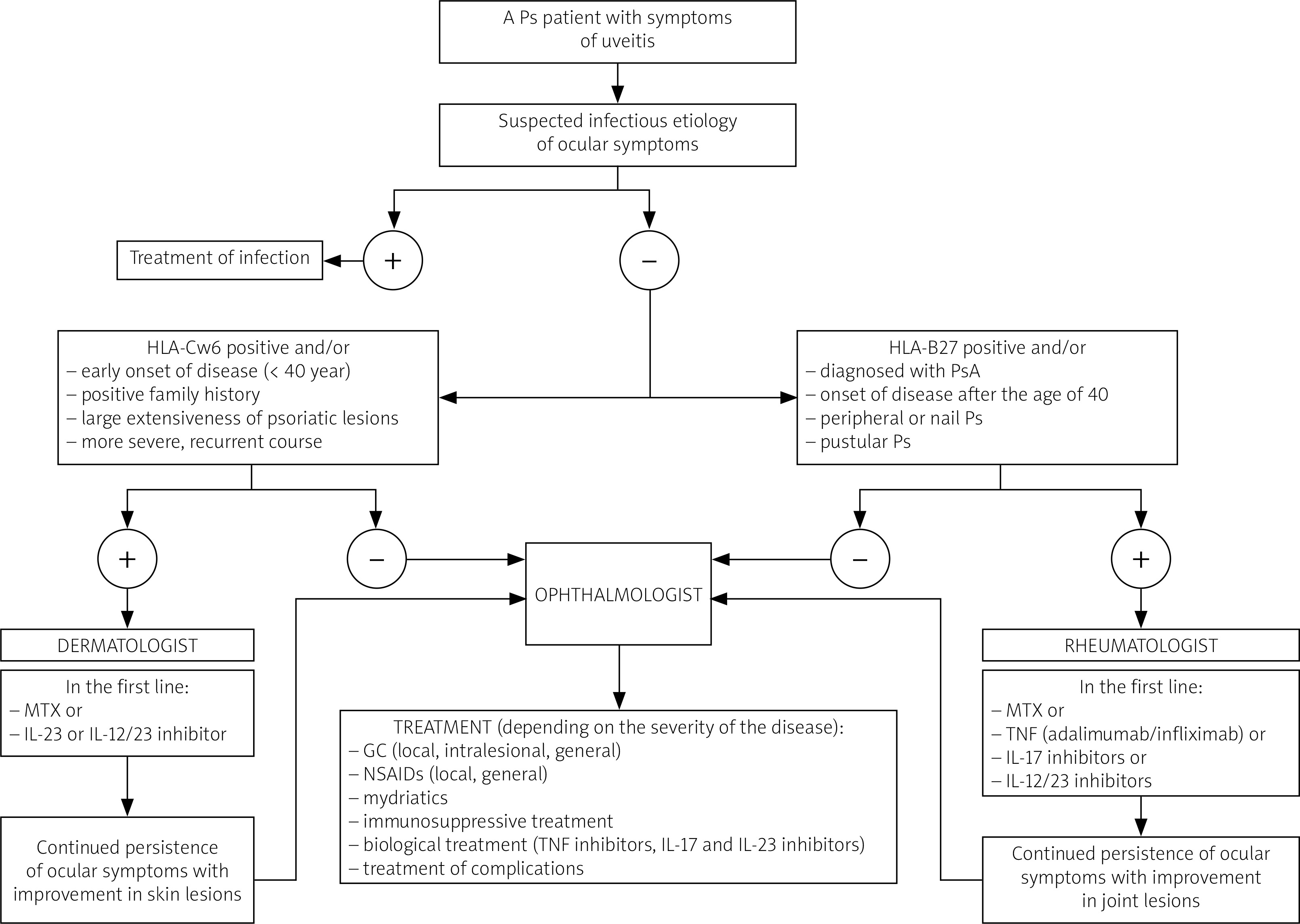
Due to the complex pathogenesis of Ps and PsA, determined by multiple genetic factors with widely documented disease conditioning, some patients may benefit from genetic testing, especially patients with uveitis, as it is often genetically determined. In particular, in differential diagnosis, it is worth considering testing for the presence of the HLA-Cw6 and HLA-B27 antigens, due to the fact that carriers of these antigens are characterized by a specific genetic phenotype that determines the clinical manifestation and course of the disease, as well as the response to the implemented treatment.
Human leukocyte antigen Cw6 is a genetic biomarker that predisposes to the development of skin manifestation of Ps (20× higher risk) and determines the course of the disease. Patients with HLA-Cw6 have a greater predisposition to type I Ps (the gene is present in 80% of patients with type I and about 15% with type II), which is characterized by: 1) early onset of disease (usually begins before 40 years of age, often in childhood or adolescence); 2) positive family history; 3) large extent of psoriatic lesions; 4) more severe, recurrent course; 5) worse response to treatment than type II. In these patients, plaques on arms, legs, and trunk and Koebner’s phenomenon occur more often, whereas nail and scalp involvement is observed less frequently. In addition, guttate Ps and photosensitive Ps are more common. It has been shown that in this group of patients, common triggers for Ps include streptococcal pharyngitis/tonsillitis, stress (20× increased risk), obesity (35× increased risk), and smoking. The risk of developing PsA is slightly increased in Ps patients with HLA-Cw6, but its occurrence is usually preceded by skin lesions long before joint symptoms appear (late-onset PsA). In addition, PsA in these patients correlates with a positive family history, and early and severe Ps. Moreover, HLA-Cw6 is a marker of a stronger response to MTX and biological treatment with and IL-12/23 or IL-23 inhibitor (but with no clear association with TNF and IL-17 inhibitors). Studies have also shown that HLA-Cw2 significantly increases the risk of developing PsA. In contrast, HLA-Cw4 does not appear to confer increased risk; in fact, some studies have even suggested a possible protective effect [16, 42, 72–75].
Human leukocyte antigen B27 is a strong genetic marker that predisposes to joint involvement in patients with Ps and correlates with the severity of joint manifestations in PsA. Greater predisposition to PsA is characterized by: 1) early onset of joint involvement; 2) increased risk of severe joint involvement; 3) aggressive course of the disease; 4) most often the axial form of PsA (while HLA-B38 and HLA-B39 predispose to peripheral polyarthritis); and 5) is a marker of disease poor prognosis. Patients with HLA-B27 have a greater risk of enthesitis, dactylitis, and uveitis. The HLA-B27 positive patients with a first episode of acute anterior uveitis have a higher risk of developing PsA in the future. Moreover, HLA-B27(+) Ps patients have an increased risk of early-onset PsA. The presence of the HLA-B27 antigen predisposes to higher incidence of pustular and peripheral Ps.
In addition, patients with positive HLA-B27 have increased risk of other SpA (such as ankylosing spondylitis, reactive arthritis) and inflammatory bowel disease, which may coexist with Ps. Studies have shown that HLA-B27(+) patients with uveitis respond better to TNFi (especially adalimumab, infliximab), IL-17, and IL-12/23 inhibitors [72–75].
In summary, genetic tests allow us to predict the clinical manifestation of the disease, which can help in selecting the appropriate treatment for the patient. Nevertheless, it should be noted that some patients with Ps and PsA do not have any of these genes, and this does not exclude the disease [51].
Based on the above genetic and clinical data, we propose an original diagnostic-therapeutic scheme – the DROP (Dermatologist-Rheumatologist-Ophthalmologist Pathway) – designed to facilitate the management of patients with Ps and uveitis symptoms in multidisciplinary clinical practice (Fig. 2).
Cataract
The latest studies have not revealed a statistically significant difference between the occurrence of cataract among patients with Ps and control groups [33, 36]. However, long-term use of topical or systemic GCs, anterior uveitis, increased age and metabolic diseases (e.g. diabetes) may promote cataract [29, 36–38]. Therefore, regular screening of cataract is recommended for patients, especially those undergoing long-term GC therapy. Some studies have suggested that PUVA therapy may be associated with an increased risk of ocular lens abnormalities, but other observations did not confirm these reports [76, 77].
In the case of symptoms and signs of dry eye, conjunctivitis, or blepharitis, the first therapeutic decision can be undertaken by the leading dermatologist or rheumatologist. However, in each case of ocular changes, the patient should be referred to an ophthalmologist for a full examination, final diagnosis, and initiation of targeted therapy.
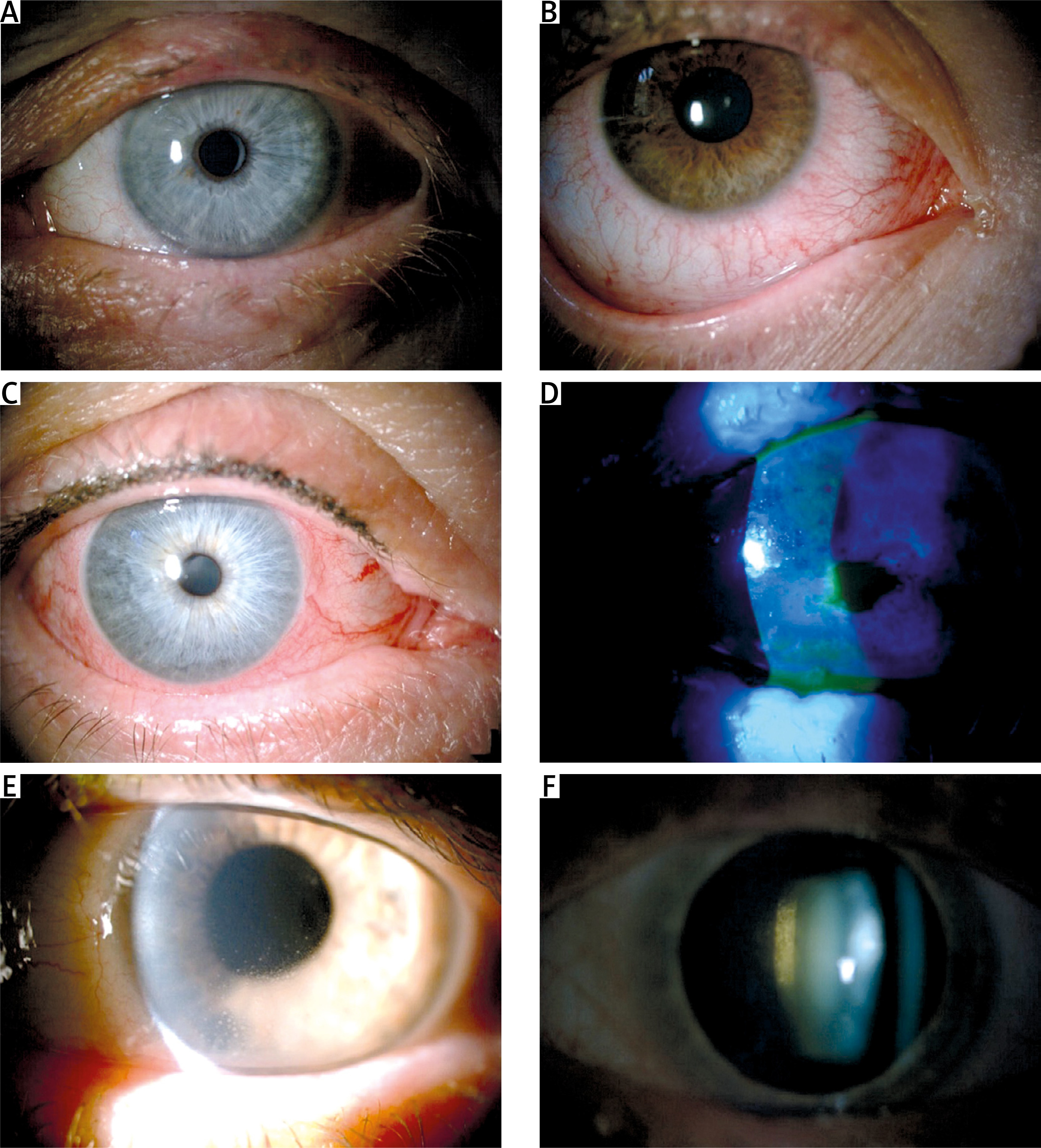
Table III presents the main symptoms, basic symptoms, and advanced treatment of the ophthalmic symptoms in Ps and PsA. Figure 3 presents pictures of the discussed ocular changes.
Table III
Ocular problems in Ps and PsA: signs and symptoms, basic and advanced treatment [12, 46, 53, 67, 76–78]
Fig. 3
Photographs of discussed ocular changes in Ps: A) blepharitis; B) conjunctivitis; C) dry eye syndrome; D) corneal epitheliopathy; E) anterior uveitis; F) cataract.
To sum up: ocular complications affect a significant percentage of patients with Ps and PsA. Their occurrence correlates with Ps activity, PASI score, and nail involvement, but is not significantly associated with disease duration. It means that they may be present even among newly diagnosed patients [33, 34, 36].
Ocular problems can affect every part of the eye and the most common manifestations include blepharitis, DED and conjunctivitis. A separate and widely described topic is uveitis in the course of PsA and, more broadly, SpA as an indicator of active disease, often associated with HLA-B27 and the need for biological treatment [24, 25, 29, 46, 53, 78].
These conditions can significantly worsen patient quality of life and lead to serious complications that may endanger eyesight. It is worth mentioning that many patients do not complain of ocular discomfort [29, 34]. This may be a consequence of focus on skin conditions or joint symptom reduction, causing symptoms concerning other organs to be underestimated or assumed not to be associated with Ps.
In patients with Ps and PsA, periodic ophthalmological check-ups should be considered, even in asymptomatic patients, due to the often insidious onset of the disease, as well as the chronic and recurrent nature of the lesions, which may lead to irreversible complications that can be prevented with early diagnosis and selection of appropriate treatment tailored to the source of the problem.
As a practical guideline, dermatologists and rheumatologists should therefore ask patients about ocular symptoms at every visit, including pain, foreign body sensation, periodic or persistent eye redness, light sensitivity, blurred vision, and visual field disorders. In physical examination, attention should be paid to the presence of eye discharge, flakes on eyelashes, conjunctival injection, and size and symmetry of the pupils. In the event of a sudden acute pain, photophobia, decreased visual acuity, eye redness or a small/irregular, minimally reactive pupil, the patient should immediately be referred to an ophthalmologist, as these conditions can seriously endanger eyesight.
Conclusions
The systemic nature of the Ps requires close cooperation of different specialists: dermatologists, rheumatologists, and ophthalmologists.
The role of the dermatologist and rheumatologist in the early diagnosis of the most common ocular complications of Ps and switching from Ps to PsA is very important. Another task of these specialists should be the patient’s education regarding the possibility of eye complications in Ps and PsA. If ocular symptoms occur, an ophthalmologic consultation should be arranged as soon as possible.
Every patient with Ps and PsA without a large area of skin involvement (PASI < 10) and without active skin involvement should undergo ophthalmologic examination annually.
Patients with PASI > 10 or those who receive systemic GCs, MTX, acitretin, phototherapy, or photochemotherapy (indicating a more active disease) should undergo ophthalmologic examination every 6 months. Those patients should be particularly closely monitored by rheumatologists and dermatologists for the occurrence of ophthalmologic symptoms.