Introduction
Spondyloarthritis (SpA) describes a group of rheumatic disorders that share a set of symptoms, including inflammatory back pain, asymmetric peripheral arthritis and enthesitis. The most frequent forms of SpA are axial SpA (axSpA) and psoriatic arthritis (PsA). Axial spondyloarthritis is characterised by progressive inflammation of the sacroiliac joints and joints of the spine, leading to bone formation and ankylosis. Its estimated prevalence is between 0.5% and 2% of the population [1, 2]. Psoriatic arthritis affects 6–41% of people with skin psoriasis [3], leading to various clinical phenotypes, including polyarthritis, oligoarthritis, dactylitis and the axial predominant type. Both axSpA and PsA are pathophysiologically associated with structural damage leading to significant disability [4]. Moreover, biomechanics might play a substantial role in the above processes, especially related to entheses [5]. Another pathology related to the skeletal system in axSpA is the loss of bone mineral density (BMD) and the deterioration of bone quality, leading to osteopenia, osteoporosis [6, 7] and vertebral fractures [8]. Although the association between SpA and osteoporosis is well established, the relationship in PsA remains insufficiently defined [9].
Current treatment guidelines in SpA focus on pharmacological and non-pharmacological approaches [10]. Pharmacotherapy aims to reduce pain and signs of inflammation by prescribing nonsteroidal anti-inflammatory drugs (NSAIDs) and immunosuppressive drugs. The latter modulate the background dysregulation of the immunological system, leading to clinical improvement. The use of various conventional synthetic, biological (bDMARDs) and targeted synthetic disease-modifying anti-rheumatic drugs is emphasized due to their high efficiency and reducing costs as so-called biosimilar medications are becoming increasingly affordable [11–13]. Additionally, the importance of exercise and appropriate physical therapy is highlighted as a complementary form of treatment [14]. Tailored exercise programmes were proven to alleviate symptoms, enhance global functioning and improve the disease course in previous studies [15–18].
Despite the suspected involvement of biomechanical stress in the pathophysiology of SpA, the underlying mechanisms and clinical implications of physical exercise on bone remodelling remain largely unidentified. Therefore, investigating and consolidating existing knowledge on the relationship between physical activity and bone remodelling in patients with SpA is of considerable clinical significance. Furthermore, given that SpA, particularly axSpA, predominantly affects younger individuals, the potential impact of mechanical stress, particularly from manual labour, may exacerbate the course of the disease [19–21]. To our knowledge, no comprehensive review addressing these factors has been published. This makes such an endeavour particularly relevant, especially in light of management approaches recommended in current clinical guidelines. Thus, the aim of this narrative review was to determine the impact of physical activity on bone remodelling and bone density in patients with SpA.
Lessons learnt from basic sciences
It is hypothesized that cascades of maladaptive bone-related processes, most prominent in entheses, are triggered by biomechanical factors. McGonagle et al. [22] proposed a model of enthesitis-based pathogenesis for SpA over 20 years ago. It underlined a possible association of repetitive compressive and shear forces occurring in entheses, in close relationship to bone and tendon, with microtrauma and activation of proinflammatory genes and healing responses. The authors emphasized that the sites of internal organ involvement in ankylosing spondylitis (AS), the radiographic subtype of axSpA (r-axSpA), e.g. the aortic root, the lung apex, the ciliary body in the eye and skin extensor surfaces are places of repetitive biomechanical forces [22].
Since that discovery, researchers have been examining the correlation between mechanical stress and SpA with regard to its potential role in the pathogenesis of the disease and its impact on disease-associated factors, including disease activity, physical functionality, pain and radiographic damage [15–17, 23]. The outcomes so far are diverse, and the impact of mechanical stress on immune system interactions and bone remodelling seems to have both positive and negative effects simultaneously [24].
Jacques et al. [5] performed an important basic science study concerning whether mechanical stress triggers inflammation and new bone formation in SpA. By molecular modification of tumour necrosis factor (TNF) production, mouse models of SpA-like arthritis were obtained. Histopathological examination of tissue specimens resulted in a conclusion that early signs of inflammation were present at entheses, eventually spreading out into the synovium. Moreover, it was found that a lack of mechanical stress prevented the development of signs of inflammation, measured both clinically and histopathologically.
In 2008, McGonagle et al. [25] confirmed a physiological reaction to repetitive biomechanical stress by discovering the formation of small spurs in healthy individuals in microanatomic studies of cadaveric entheses. In this study of SpA patients with enthesitis using ultrasound (US), McGonagle et al. [25] proved that bone erosions and osteoproliferation are distinct anatomically and temporally. He noted that erosions and new bone formation are linked to biomechanics since erosions occurred in areas of compression, while spur formation took place in regions where tensile forces are likely to be greater.
In a 2014 animal study, Jacques et al. [5] proved that mechanical load is necessary for disease onset and correlates with arthritis and new bone progression. In tail-suspended TNF delta AU-rich element (TNFΔARE) mice (with chronic and deregulated TNF production), clinical signs of arthritis in the hind paws were not observed on clinical examination, and inflammatory changes on histological examination were minor. In collagen antibody-induced arthritis tail-suspended mice, osteophytes were markedly smaller on radiological and histopathological evaluation.
The author corroborated the role of stromal cells in the initiation of enthesitis. In mature T-cell-deficient mouse models [recombination activating gene 1 (RAG-1) deficient TNFΔARE], the incidence of enthesitis was similar to that in the control group, which indicates that both T-cell-dependant and -independent mechanisms lead to enthesitis [5].
Conclusions drawn from clinical research
The evaluation of SpA imaging measures supports the hypothesis of biomechanics-related osteoproliferation in SpA. Tan et al. [19] analysed the spatial distribution of spine syndesmophytes in r-axSpA. He found that syndesmophytes occur and first develop along the vertebral rim, a place exposed to mechanical forces. Therefore, this particular occurrence suggests a possible role of biomechanics in the development of syndesmophytes in axSpA. Moreover, Tan et al. [26] observed syndesmophytes, which were less common and smaller and were located at the thoracolumbar vertebral rim near the aorta. This observation added further evidence that not only inflammation but also biomechanical forces extrinsic to the spine play a role in new bone formation in axSpA. A key topic in many studies is the presence of bone marrow oedema (BME) in sacroiliac joints (SIJ) on magnetic resonance imaging (MRI). The bone marrow oedema is hallmark of axSpA, but it may also be present in healthy individuals such as postpartum women, athletes and others [27, 28]. The bone marrow oedema is often present in SIJ exposed to high biomechanical forces found in postpartum women, patients with disc herniation, athletes (runners, hockey players), military recruits and professionals, which suggests an impact of mechanical stress [27, 28]. Although in the study by Varkas et al. [29], no differences were observed in SIJ MRI findings in military recruits after 6 weeks of intense physical training, it is noteworthy that a high prevalence of MRI lesions (40.9%) was already present in this population before the training began.
In studies investigating the correlation between the presence of syndesmophytes in patients with r-axSpA, associations were found with disease duration, patient age, and elevated levels of osteocalcin (a marker of osteoproliferation), whereas no association was observed with disease activity or low BMD [30, 31].
Obesity has been associated with higher disease activity and with axial and peripheral osteoproliferation and entheseal inflammation, as reported by Maas et al. [32] and Bakirci et al. [33]. Although involved in immunological reactions [34], obesity increases biomechanical forces in axial and peripheral joints.
Bone density in spondyloarthropathies
Low BMD is already present in 47% of patients with early SpA (with a median disease duration of 6 years), including osteoporosis in 9–18% [7, 9]. It is associated with male sex, in contrast to the healthy population, as well as with high disease activity and decreased functional performance [7, 31]. However, in PsA, osteopenia and osteoporosis appear to occur with a prevalence similar to that of the general population [9]. Current studies underline the role of proinflammatory cytokines in bone resorption and the pathogenesis of low bone mass in SpA [35]. Studies on bone turnover markers indicate that the level of N-terminal telopeptide – a marker of bone resorption – in patients with r-axSpA is associated with disease activity, C-reactive protein levels, low BMD, and the occurrence of fractures, while it is not associated with the presence of syndesmophytes [30].
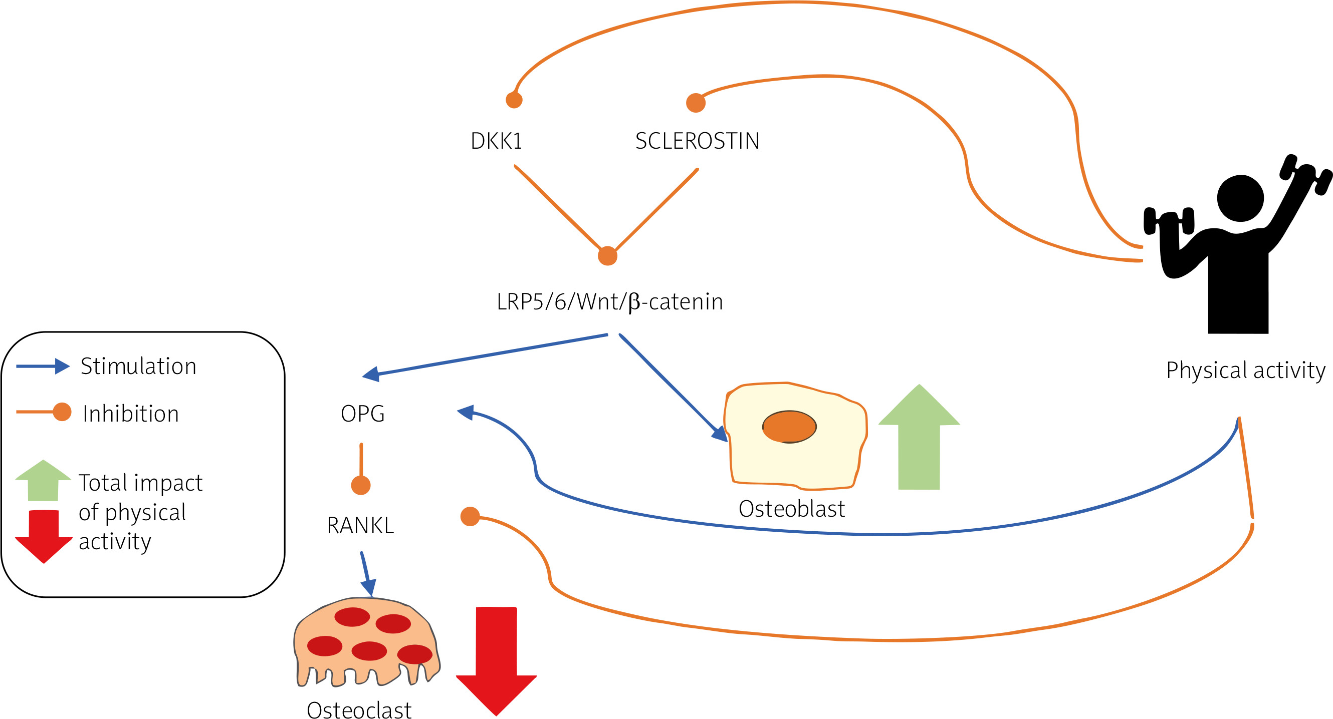
Inflammatory cytokines elevated in SpA, such as TNF and interleukin-17, impact bone remodelling. Under physiological conditions, inflammation, bone resorption, and formation are essential for tissue healing. In pathological conditions, however, these processes contribute to low BMD, osteoporosis, syndesmophyte formation, and ankylosis. Several osteoimmunological pathways play a role in r-axSpA, including bone morphogenetic proteins (BMPs), the Wnt signalling pathway, and Hedgehog signalling (Hh) [36]. A fundamental pathway in bone metabolism is Wnt. In the presence of inflammation, TNF promotes the expression of Dickkopf-1 (DKK1) and sclerostin (Wnt inhibitors), inhibits osteoblast differentiation, and enhances bone resorption by increasing the receptor activator of nuclear factor kappa-B ligand (RANKL)/osteoprotegerin (OPG) ratio [36].
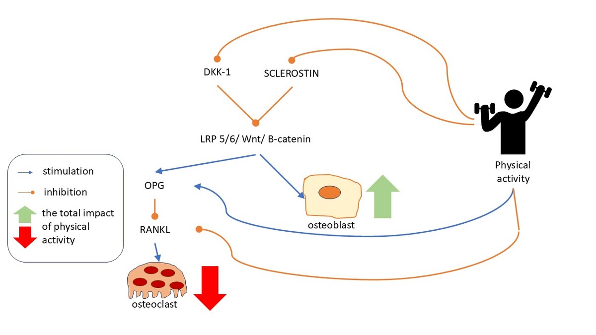
Physical activity acts in the opposite manner: it reduces the expression of DKK1, sclerostin, and RANKL; increases the expression of OPG; and overall promotes osteoblast proliferation and activity while inhibiting osteoclast differentiation and activation [37, 38].
Molecular mechanisms of the effect of physical activity on bone metabolism are shown in Figure 1.
Fig. 1
Molecular mechanisms underlying the effects of physical activity on bone metabolism [37, 38].
DKK1 – Dickkopf-1, LRP5/6 – low-density lipoprotein receptor-related protein 5 and 6, OPG – osteoprotegerin, RANKL – receptor activator of nuclear factor κ-B ligand, Wnt/β-catenin – Wnt/β-catenin signalling pathway.
Tumour necrosis factor blockers in r-axSpA lower DKK1 and BMP 7 levels [39], decrease expression of Indian Hh [40], and increase the OPG/sRANKL ratio [41]. Several meta-analyses have demonstrated that anti-TNF agents improve BMD [42, 43].
The effect of antiresorptive drugs on bone metabolism in r-axSpA remains insufficiently explored. To date, no double-blind randomized clinical trial evaluating the impact of bisphosphonates or RANKL inhibitors on BMD in SpA patients has been conducted. In a meta-analysis comparing outcomes of r-axSpA patients treated with and without bisphosphonates, published between January 2000 and March 2020, only 6 comparative studies were identified [44]. According to this meta-analysis, bisphosphonates do not improve lumbar or femoral BMD or disease activity in r-axSpA; however, limitations such as small sample sizes and short follow-up durations should be taken into consideration [44].
In spondyloarthritis, bone loss is driven by inflammation, which promotes increased osteoclastogenesis through both RANKL-dependent and -independent pathways, while simultaneously limiting osteoblast formation [45]. Thus, antiresorptive therapies, such as bisphosphonates and RANKL inhibitors, may be expected to improve bone mass in patients with low BMD and active inflammation.
Studies investigating the effects of denosumab in SpA are scarce. A South Korean pilot study demonstrated the efficacy of denosumab in improving lumbar and total hip BMD, without affecting disease activity or spinal new bone formation [46]. One case series reported an increase in BMD, measured by quantitative computed tomography, after 1 and 2 years of denosumab treatment [47]. Large, long-term randomised clinical trials evaluating the effect of denosumab in SpA are required. Considerably more data are available regarding the efficacy of denosumab in rheumatoid arthritis: a meta-analysis of 6 randomised controlled trials showed that denosumab improves BMD and prevents joint erosions in patients with osteoporosis and rheumatoid arthritis [48].
Numerous clinical studies have shown that physical activity increases peak bone mass and reduces the risk of osteoporosis; therefore, it is recommended for both its treatment and prevention in the general population [49]. Given the inflammatory nature of osteoporosis in SpA, it is important to further explore the effects of physical activity on both disease activity and bone density in this population.
Methods
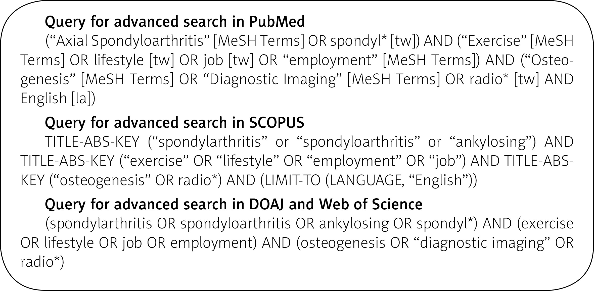
This narrative review article was conducted in accordance with methodology provided by expert guideline articles, including a manuscript by Gasparyan et al. [50]. Also, to improve the quality of the obtained results, the validity of the methodology and possible reproducibility of this study, the article selection process followed the PRISMA 2020 statement. The PubMed, Scopus, Directory of Open Access Journals, and Web of Science bibliographic databases were searched. Search terms included Medical Subject Headings terminology and additional concepts to obtain the maximum number of results. Specific search queries used in the process are provided in the appendix. The initial data extraction, including article titles and authors’ names, was performed on 20 May 2024. The initial search was repeated in October 2024 and shortly before submission of this article (May 2025), to provide the most up-to-date information regarding the examined phenomena. The articles were then screened by 2 authors (AK, MW), according to the provided literature selection flowchart. The search strategy is presented in Figure 2.
The following inclusion criteria were applied:
studies conducted on adult patients (over 18 years of age);
participants meeting the appropriate classification criteria, relevant to the aim of the study (the 1984 New York criteria for the r-axSpA (AS), the 2010 ASAS criteria for the non-radiographic form of SpA and the 2006 CASPAR criteria for PsA) [51–53];
studies comparing the assessed indicators (imaging studies or bone density assessment) in populations with high or low physical activity (recreational or occupational);
randomized controlled trials (RCTs), non-RCTs, randomized uncontrolled trials, or uncontrolled studies (e.g. before vs. after trials), cross-sectional studies, observational studies, including prospective and retrospective studies;
studies in the English language;
full-text manuscript available.
Studies were excluded if:
the study scope was outside the aim of the current analysis (e.g. physical activity was not assessed, or the study focused on the impact of physical activity solely on indicators other than imaging or bone density, such as range of motion, functionality, disease activity, quality of life, etc.);
the diagnostic criteria and selection of patients were inadequate;
the risk of biased results, due to methodological issues, was considerable, as assessed by 2 authors (AK, MW);
the full-text manuscript was unavailable or was in a language other than English.
Also, other review articles (systematic analyses, meta-analyses), case studies, study protocols, congress abstracts, editorials and letters to the editor were not included in the current analysis. No restrictions were applied regarding the date of publication. If uncertainty arose considering the inclusion of a specific article, it was further discussed among all authors, leading to a mutual decision.
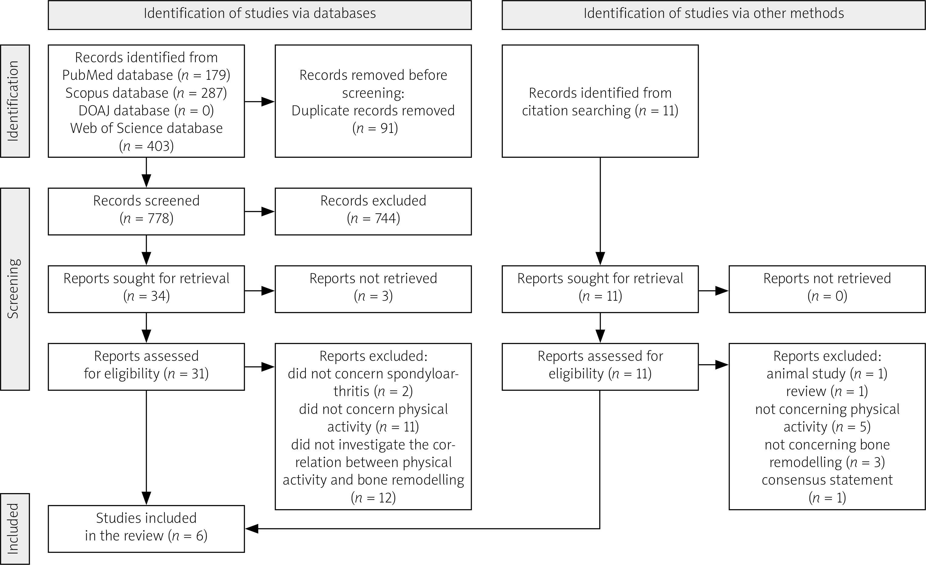
After completing the search on the impact of physical activity on radiological progression in patients with SpA, 2 researchers selected articles in the following stages: removal of duplicates, review of titles and abstracts, review of full texts and review of the references and citations of selected articles. We also included additional papers recommended by all co-authors, deemed significant for the context of this review. The final reference list was compiled based on their relevance to the key concepts the authors intended to emphasize in the manuscript. The article selection process is presented in Figure 3.
Fig. 3
Article selection process according to the PRISMA 2020 flow diagram.
DOAJ – Directory of Open Access Journals.
From the included articles, the following data were systematically extracted: study population (type of SpA, disease duration, use of biologic treatment), study type, type of physical activity (physical work, exercises) and results of radiological studies.
Results
The initial and additional database searches found 869 articles: 179 articles in PubMed, 287 in Scopus, 403 articles in Web of Science, and no article was identified in Directory of Open Access Journals. After removing duplicates, 778 articles remained. Title and abstract screening left 34 articles that qualified for full-text analysis. After full text analysis, 6 eligible articles remained. The search was further expanded by citation screening and reference screening, and no more articles were included in the final analysis.
Despite searching 4 major bibliographic databases, using liberal search criteria, and having the searches conducted by 2 team members, to the authors’ best knowledge, at the time of the additional search (May 2025), no studies assessing the impact of physical activity on bone density in patients with SpA were conducted.
The following research concerned the impact of physical activity on bone remodelling in axSpA patients: after searching 4 databases, using liberal search criteria, and searches being conducted by 2 team members, only 6 articles met the inclusion criteria and were analysed.
Characteristics of included studies
The analysed populations varied significantly. Three studies included patients with PsA, including one with axial PsA, while the remaining three involved patients with r-axSpA. The characteristics of the patients also differed significantly. Three studies focused on early SpA (2 on PsA and 1 on r-axSpA), with a median disease duration of 3–6 years [54–56], whereas three other studies examined longstanding SpA (2 on r-axSpA and 1 on PsA), with a mean disease duration ranging from 11 to 32 years [19–21]. Notably, the proportion of patients treated with bDMARDs differed across studies. In 1 study, none of the patients received bDMARD treatment at baseline, whereas in the others, this percentage ranged from 27% to 48%. Among the analysed studies, only 1 provided information about steroid therapy; moreover, not all of them reported on the use of NSAIDs or synthetic DMARDs. In the study by Ward [19], glucocorticosteroids were used in only 7% of patients [57–59].
Two of the included studies were randomized controlled trials exploring the impact of high-intensity interval training (HIIT) on numerous outcomes, including radiographic structural damage, among PsA patients. Thomsen et al. [56] assessed the impact of 11 weeks of HIIT on imaging of the spine, peripheral joints and entheses in MRI (axial) and US (peripheral) compared to the control group. The analysed cohort comprised early PsA patients, and 31% of them were treated with bDMARDs. Similarly, Chronaiou et al. [55] analysed the impact of 11 weeks of HIIT on MRI of the spine in early axial PsA patients compared to the control group. In both studies, early inflammatory changes of the spine were scored using the Spondylarthritis Research Consortium of Canada (SPARCC) BME score, and inflammation of peripheral joints and entheses was assessed using semiquantitative gradation of brightness mode and Power Doppler images from 0 to 3. In the study by Chronaiou et al. [55], the images were scored by 1 reader in radiological evaluation for BME using the Assessment in SpondyloArthritis International Society criteria, by another reader in SPARCC score and by extraction of textural features.
Slouma et al. [54] investigated a correlation between levels of physical activity measured by 2 scales, the University of California and Los Angeles activity scale and the Tegner activity scale, supplemented with information regarding the weekly duration of exercise and physical activity, and disease progression as assessed by US imaging of 14 predefined entheses in patients with early r-axSpA. Ultrasound imaging was scored using the Spanish Enthesitis Index, Glasgow Ultrasound Enthesitis Scoring System and Madrid Sonographic Enthesitis Index.
The remaining 3 studies analysed the association between professional work and radiographic structural damage. Participants were categorized by their occupational activity into 2 groups: blue-collar workers with physically demanding jobs and white-collar workers with physically undemanding jobs. The rationale for this approach is that physically demanding jobs are presumably associated with more mechanical stress to the spine than sedentary ones. In a study by Ramiro et al. [21], the job type was determined by consensus, and Zhou et al. [20] and Ward et al. [19] used the Occupational Information Network (job classification database) to determine specific attributes of jobs (such as workers’ abilities, measures of work context, etc.) and found associations between particular activities and radiographic progression. The included studies involved participants with longstanding r-axSpA [19, 21] and PsA [20]. Radiograms were taken every 2 years in the Ramiro et al. [21] and Zhou et al. [20] studies and once in the study by Ward et al. [19] Cervical and lumbar spine X-rays were scored using the modified Stoke Ankylosing Spondylitis Spine Score (mSASSS) [21, 20] or the Bath Ankylosing Spondylitis Radiology Index for the spine (BASRI-s) [19]. Peripheral joint radiograms were scored using the modified Steinbrocker score [20]. Of concern, in the study by Ward et al. [19], the images were scored by only 1 reader.
Both scoring systems, mSASSS and BASRI-s, rate the degree of structural damage (including erosions, sclerosis or fusion) in the cervical and lumbar spine. The Bath Ankylosing Spondylitis Radiology Index for the spine additionally rates damage to SIJ. Detailed characteristics of the included studies are presented in Table I.
Table I
Characteristics of included studies
| Author, year, country, reference | Disorder | Study design | Study population | Disease duration (years) | bDMARDs | csDMARDs | GCs | NSAIDs |
|---|---|---|---|---|---|---|---|---|
| Thomsen et al. 2023, Norway [56]* | PsA | RCT | 67 | 3–5.5 (median) | 31% | 85% | No data | No data |
| Chronaiou et al. 2022, Norway [55, 57]* | axPsA | RCT | 39 | 4–6 (median) | 33% | 87% | No data | No data |
| Ramiro et al. 2015, Netherlands/Belgium/ France [21, 58] | r-axSpA | Cross- sectional | 184 | 11 (mean) | 0% at year 0, 29% at year 12 | No data | No data | 68% |
| Zhou et al. 2019, Canada [20] | PsA | Cross- sectional | 307 | 21 (mean) | 48% (ever) | 76% (ever) | No data | 92% (ever) |
| Ward et al. 2008, USA [19, 59] | r-axSpA | Cross- sectional | 397 | 31.9 (mean) | 27% | 24% | 7% | 83% |
| Slouma et al. 2024, Tunisia [54] | r-axSpA | Cross- sectional | 37 | 5.3 (median) | 27% | No data | No data | 92% (32% continuous) |
* Thomsen’s and Chronaiou’s studies are based on a common cohort of patients recruited from St. Olavs Hospital and Norwegian University of Science and Technology, Trondheim, Norway, between 2013 and 2015.
axPsA – axial psoriatic arthritis, bDMARDs – biological disease-modifying anti-rheumatic drugs, csDMARDs – conventional synthetic disease-modifying anti-rheumatic drugs, GCs – glucocorticosteroids, NSAIDs – nonsteroidal anti-inflammatory drugs, PsA – psoriatic arthritis, r-axSpA – radiographic axial spondyloarthritis, RCT – randomized controlled trial.
Outcomes of the analysed studies
Detailed information about the results of the analysed studies is presented in Table II.
Table II
Results of analysed studies
| Author, year, country, reference | Physical activity type | Intervals of measurements | Instruments used | Scales |
|---|---|---|---|---|
| Thomsen et al. 2023, Norway [56] | Intervention group: HIIT for 11 weeks; control group: not to change their physical exercise habits | At baseline and at 3-month follow-up | MRI of the SIJ and the spine (C, Th, L), US of the peripheral joints and entheses (mandatory: 34 joints, 10 entheses and additional swollen or tender) | SPARCC-BME score of the SIJ and spine; B-mode and PD signals in joints and entheses were semi-quantitatively graded 0–3 |
| Conclusions | ||||
| No evidence of an increased risk of inflammation. | ||||
| Chronaiou et al. 2022, Norway [55] | Intervention group: HIIT for 11 weeks; control group: not to change their physical exercise habits | At baseline and at 3-month follow-up | MRI of the spine (C, Th, L) | SPARCC |
| Conclusions | ||||
| No significant changes | ||||
| Ramiro et al. 2015, Netherlands/Belgium/France [21] | Job type | 2-year intervals | X-rays of the spine (C, L) | mSASSS |
| Conclusions | ||||
| Physically demanding jobs may amplify the potentiating effects of inflammation on bone formation in AS | ||||
| Zhou et al. 2019, Canada [20] | Job type | 2-year intervals | X-rays of the spine (C, L) and peripheral joints (hands and feet) | mSS and mSASSS |
| Conclusions | ||||
| Repetitive hand movements and finger dexterity were independently associated with peripheral radiographic damage. None of the occupational exposures were significantly associated with axial damage | ||||
| Ward et al. 2008, USA [19] | Job type | One time | X-rays of the spine and the pelvic | BASRI-s |
| Conclusions | ||||
| Those whose past jobs required more dynamic flexibility (the ability to repeatedly bend, stretch, twist, or reach), extensive flexibility and exposure to whole body vibration had significantly higher BASRI-s scores | ||||
| Slouma et al. 2024, Tunisia [54] | UCLA activity scale, Tegner activity scale, patient’s estimated duration of exercise and of physical activity weekly | One time | Ultrasound of 14 entheses in each patient | SEI, GUESS, MASEI, presence of enthesophyte, hypoechogenicity, calcification, or bony erosion |
| Conclusions | ||||
| Lack of a significant difference in enthesis ultrasound characteristics between patients practising or not practising exercise. Patients performing exercise had a lower risk of having ≥ 3 ultrasonographic enthesophytes. | ||||
[i] BASRI-s – Bath AS Radiology Index for the spine, BME – bone marrow oedema, GUESS – Glasgow Ultrasound Enthesitis Scoring System, HIIT – high-intensity interval training, MASEI – Madrid Sonographic Enthesitis Index, MRI – magnetic resonance imaging, mSASSS – modified Stokes Ankylosing Spondylitis Spine Score, mSS – modified Steinbrocker score, SIJ – sacroiliac joints, SPARCC – Spondyloarthritis Research Consortium of Canada, SEI – Spanish Enthesitis Index, UCLA – University of California and Los Angeles.
Briefly, in a study by Thomsen et al. [56], the authors concluded that there is no significant influence of HIIT on the severity of inflammation in the peripheral joints, entheses, SIJ and spine assessed by US and MRI.
In an analysis by Chronaiou et al. [55], the number of participants with BME changes on radiological evaluation, in SPARCC scores or in textural features did not differ significantly between the 11-week HIIT group and the control group.
Slouma et al. [54] found no significant differences in US image of entheses in patients with r-axSpA between individuals practising and not practising exercise. Moreover, in the multivariate analysis, they found a lower occurrence of 3 or more enthesophytes in patients who exercised.
Ramiro et al. [21] concluded that patients with high mSASSS are mainly men, blue-collar workers and smokers. They suggested that blue-collar jobs amplified the effect of disease activity on radiographic damage in comparison with white-collar jobs.
Zhou et al. [20], analysing 307 patients with longstanding PsA, found a direct association between damage to the peripheral joints and prolonged repetitive hand movements and a high level of finger dexterity. No correlation was found between mSASSS and occupation-related mechanical factors.
Ward et al. [19] found that occupational activities, such as bending, twisting, reaching and stretching, and exposure to whole-body vibration are associated with greater radiographic damage in axial joints.
Discussion
The authors of the analysed studies reached different conclusions concerning the impact of physical activity on the clinical progression of SpA. Researchers assessing the role of HIIT found no significant changes in inflammation of the spine, peripheral joints and enthesis on MRI and US [54–56]. However, concerning these results, a few methodological issues appear. The relatively short follow-up period of 11 weeks might not be sufficient to reveal an impact on bone, joint and entheses structures. The remodelling processes are time consuming; thus, a longer follow-up study could improve the validity of the obtained results. On the other hand, the authors of studies exploring the influence of occupational activity type demonstrated an association between job type and radiographic structural damage. However, these cross-sectional studies can only draw conclusions about possible correlations, not potential causality.
Many questions remain unanswered, and thus high-quality, prospective interventional studies are necessary. However, developing such studies poses several challenges. Spondyloarthritis is a heterogeneous condition, and different stages of the disease may impact bone remodelling in various ways, making it difficult to standardize the study population. Moreover, the various forms of spondyloarthropathies differ from one another in their bone metabolism. Despite some common pathophysiological mechanisms, in contrast to r-axSpA, in PsA osteoporosis does not seem to be a significant comorbidity [9]. Bone remodelling in SpA involves both new bone formation (e.g. syndesmophyte growth, juxta-articular bone formation, ankylosis) and bone loss (e.g. osteoporosis, erosions), and physical activity may affect these processes differently, complicating the measurement of clear outcomes related to bone tissue [6, 7, 36]. The type, intensity and duration of physical activity can vary significantly between individuals, making it challenging to design a regimen that is both feasible for enrolled participants and possible to capture in imaging measurements.
Furthermore, factors such as medication use (e.g. DMARDs, NSAIDs, glucocorticosteroids, antiresorptive drugs), diet and vitamin D levels also affect bone health in SpA, making it difficult to isolate the impact of physical activity alone. Furthermore, some studies suggest that vitamin D supplementation may improve disease activity and, in doing so, influence bone metabolism by reducing inflammation [60, 61]. It may take months or even years to capture a least significant change in bone remodelling, especially in axSpA, necessitating long-term follow-up, which increases study cost, complexity and the risk of participant dropout [62]. Measuring bone remodelling accurately is another challenge. Imaging methods such as MRI or dual-energy X-ray absorptiometry (DXA) may not detect early or subtle changes in bone tissue, and biomarkers of bone turnover can be variable and influenced by several irrelevant factors [63]. Although DXA is the gold-standard method for fracture risk assessment, the presence of syndesmophytes may lead to false-negative results, making it unsuitable for axSpA patients. Nevertheless, there is currently no other widely available and recommended alternative [64]. Additionally, ethical considerations may arise in designing studies that prescribe or limit physical activity, particularly for patients with more severe forms of SpA. Recruiting participants who are willing and able to adhere to a specific physical activity protocol can also be challenging [65]. Moreover, the assessment of physical activity may be imprecise, particularly as patients tend to overestimate the duration of their physical activity [66].
The current narrative review article has several strengths. It addresses a clinically relevant topic with potential implications for clinical practice. Currently, clinicians managing patients with axSpA complicated by osteoporosis adhere to osteoporosis treatment guidelines developed for the general population. In the context of increasing availability and more widespread use of biologic therapy – known to have a positive effect on bone mass – there is a clear need for studies evaluating the effectiveness of antiresorptive agents, DMARDs, and combined therapeutic approaches.
In our research an extensive search process, including citation and reference screening of 4 major bibliographic databases, covered relevant studies. A rigorous methodological approach also enhanced the validity of the findings. However, the study also has a few limitations.
Despite the thorough search process, only a few articles were ultimately included. There is also a risk of subjective author bias. Furthermore, due to the nature of the studies and the limited number of suitable articles, it was not possible to perform a quantitative synthesis.
In conclusion, the impact of physical activity on bone remodelling in SpA warrants greater attention due to its important clinical implications, which could contribute to overall patient health. However, it is important to recognize that significant knowledge gaps remain, and further high-quality, prospective research is crucial to address these gaps and deepen our understanding of this complex relationship.