Introduction
Systemic sclerosis (SSc) is a complex autoimmune disease whose pathogenesis is based on 3 pillars: vasculopathy, immune system dysregulation, and progressive fibrosis of the skin and internal organs [1–3]. Vascular abnormalities initiate the process through endothelial dysfunction and microvascular rarefaction, leading to chronic tissue hypoxia. This environment triggers immune dysregulation, characterized by the activation of both innate and adaptive immunity. B-cells play a pivotal role here, not only by producing specific autoantibodies (e.g., anti-topoisomerase I [anti-Scl-70]) but also by secreting pro-fibrotic cytokines such as interleukin-6 (IL-6) and transforming growth factor-β (TGF-β). This persistent inflammatory signaling drives fibroblast differentiation into myofibroblasts, leading to excessive extracellular matrix deposition and progressive organ fibrosis [1, 3, 4]. Despite the availability of immunosuppressive drugs (such as cyclophosphamide or mycophenolate mofetil), many patients do not achieve sustained remission, and the disease continues to be associated with significant morbidity and mortality [2, 4]. The prognosis in severe forms of the disease remains poor, and traditional treatment rarely leads to long-term remission without the need for continuous drug administration [1].
Autologous hematopoietic stem cell transplantation (HSCT) has become an alternative for patients with severe, treatment-refractory SSc. Studies such as ASTIS and SCOT have demonstrated the superiority of HSCT over cyclophosphamide, showing that it can induce deep and durable remission. Long-term observations confirm that HSCT significantly reduces skin fibrosis and stabilizes organ function, with 5-year overall survival (OS) rates ranging from 79% to 96%. However, this procedure remains associated with a high risk of peri-procedural toxicity and mortality [4, 5]. Specifically, treatment-related mortality associated with HSCT can reach up to 10% in some cohorts, primarily due to cardiac toxicity or infectious complications. This significant safety concern, alongside the rigorous eligibility criteria, limits its application to a highly selected group of patients. In this context, chimeric antigen receptor (CAR) T-cell technology, originally developed for hematological malignancies, has emerged as a potential therapeutic strategy.
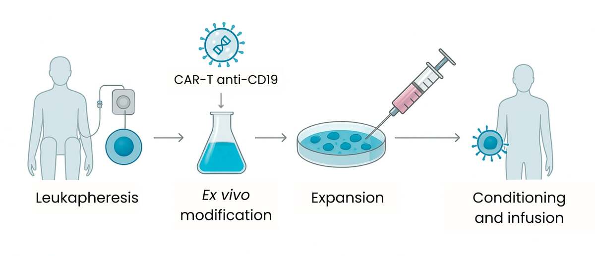
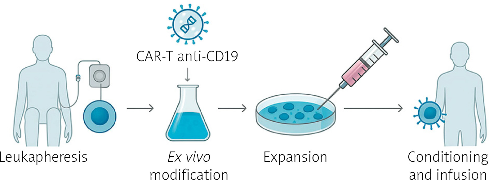
The CAR is a modular synthetic fusion protein designed to redirect T-cell specificity toward a predefined surface antigen [4–6]. The clinical application of autologous CAR-T cells involves a complex “vein-to-vein” manufacturing process, encompassing leukapheresis, ex vivo genetic engineering, and a lymphodepletion conditioning regimen (typically comprising fludarabine and cyclophosphamide) to promote CAR-T cell expansion [5, 6]. Therapy with CAR-T targeting the CD19 antigen (CD19-CAR-T) aims at deep B-cell depletion, which may restore immunological tolerance [1, 6]. Recent reports, including case series, indicate that this therapy is feasible and may lead to clinical symptom resolution in SSc patients refractory to standard treatment [1, 7]. In addition to classic autologous CAR-T preparations, allogeneic (“off-the-shelf”) products, with genetic modification (e.g., using CRISPR/Cas9), are also being investigated to reduce the risk of rejection, thereby increasing the accessibility of this method [8].
Material and methods
This manuscript is a narrative review without formal quality assessment and aims to provide a comprehensive, clinically relevant synthesis of CAR-T cell therapy approaches in SSc. The literature was reviewed by searching the scientific databases PubMed and Scopus. The materials presented at the most significant international rheumatology congresses, such as the European Alliance of Associations for Rheumatology (EULAR) and the American College of Rheumatology (ACR), were also analyzed. To ensure retrieval of all relevant literature, the search strategy combined free-text terms with Medical Subject Headings (MeSH). The search terms included the following combinations of keywords: (“CAR-T” OR “CD19-CAR-T” OR “CD19 CAR-T” OR “chimeric antigen receptor” OR “chimeric antigen receptor T-cell” OR “CAR-T cell therapy”) AND (“scleroderma” OR “systemic sclerosis” OR “SSc pathogenesis” OR “SSc”). MeSH terms were used to enhance search precision, including “Scleroderma, Systemic” and “Receptors, Chimeric Antigen.”
The inclusion criteria were as follows:
original scientific articles (clinical trials and observational studies) on the efficacy or safety of CAR-T cell therapy in SSc,
studies published between January 2023 and January 2026.
The exclusion criteria included studies that:
lacked full text,
were non-English publications,
described the use of CAR-natural killer (NK) cell therapy.
The initial stage of the process involved selecting articles based on their titles. In the second stage, the importance of the abstract was considered, and finally, the full texts were examined. The reference lists of the included studies and relevant reviews were also screened to identify any additional records. Two independent reviewers screened the articles for relevance to the review’s objectives following a database search. This review included 21 patients with SSc who had undergone CAR-T cell therapy. Auth et al. [7] reported a case series involving 6 patients with diffuse SSc (dSSc). Notably, this series incorporated preliminary data from 2 patients with SSc enrolled in the CASTLE study (NCT06347718), a phase 1/2a basket trial evaluating the safety and efficacy of zorpocabtagene autoleucel. Consequently, the present review considers 4 patients described by Auth et al. [7] and 9 patients from the CASTLE study. Additionally, 5 patients from the study by Pecher et al. [9] were included. The remaining 3 patients were identified from case reports.
Discussion
Mechanistic rationale for CD19-targeted chimeric antigen receptor T-cell therapy in systemic sclerosis
The application of cellular therapy in SSc stems from the fundamental role of B lymphocytes in the initiation and perpetuation of the disease process, which is confirmed by numerous studies on SSc pathogenesis [7, 10]. B lymphocytes in SSc exhibit features of constitutive hyperreactivity, which manifests, among other things, as approximately 20% increased expression of the signaling molecule CD19 compared to healthy individuals. This CD19 overexpression lowers the activation threshold of B cells, leading to excessive stimulation and disruption of homeostasis, as manifested by the expansion of naive B lymphocytes alongside a decrease in the number of memory cells [10]. A consequence of this dysregulation is the differentiation of B lymphocytes into plasma cells that produce autoantibodies strictly correlated with the clinical phenotype, such as anti-Scl-70 and anti-RNA polymerase III (ARPA), whose titers are linked to disease activity and prognosis [7, 8]. Importantly, the pathogenicity of B lymphocytes in SSc extends beyond the production of classic nuclear autoantibodies; functional agonist autoantibodies have also been identified, directed, for example, against the platelet-derived growth factor receptor (PDGF) or the angiotensin II type 1 receptor. These antibodies can directly stimulate fibroblasts to produce collagen and promote vascular changes, thus forming a direct link between autoimmunity and fibrosis [10]. Furthermore, activated B lymphocytes perform crucial effector functions, secreting pro-inflammatory and pro-fibrotic cytokines, such as IL-6 and TGF-β, and acting as antigen-presenting cells, which stimulate T lymphocytes and drive chronic inflammation [7, 9].
Although B-lymphocyte depletion strategies using anti-CD20 monoclonal antibodies, such as rituximab (RTX), have demonstrated some clinical efficacy in improving skin conditions, their potential in SSc is limited by the incomplete elimination of pathogenic clones [6, 10, 11]. Rituximab often fails to achieve complete B-lymphocyte depletion in deep tissue compartments, such as fibrotic skin or lymph nodes, where these cells can survive and perpetuate the disease process [1, 6]. CAR-T anti-CD19 therapy offers a mechanistic advantage due to its broader target range and distinct mode of action. The CD19 antigen is expressed on a broader spectrum of the B-lymphocyte lineage than CD20, covering stages from pro-B lymphocytes through mature cells up to plasmablasts and early plasma cells [1, 12]. Plasmablasts, a key source of autoantibodies, often lack CD20 expression, rendering them resistant to RTX treatment, but they remain susceptible to anti-CD19 CAR-T therapy [1, 6, 10]. Furthermore, as a “living drug,” CAR-T cells are capable of active migration from the bloodstream into inflamed tissues, allowing for effective elimination of residual B lymphocytes inaccessible to passively acting antibodies [1, 10] (Fig. 1).
Fig. 1
Production and administration process of autologous anti-CD19 CAR-T cell therapy.
Anti-CD19 – anti-cluster of differentiation 19, CAR-T – chimeric antigen receptor T cells.
Support for this mechanism comes from histopathological studies showing extensive elimination of B lymphocytes from the skin of SSc patients following CAR-T cell infusion [10]. The complete depletion of B lymphocytes at both the peripheral and tissue levels by CAR-T cells may promote immune system reconstitution, a phenomenon commonly referred to as an “immunological reset” [1, 7]. Available clinical observations indicate that after a period of B-lymphocyte aplasia, lasting on average approximately 112 days, the B-cell population undergoes reconstitution characterized predominantly by naïve cells with a newly formed receptor repertoire lacking autoreactive clones [1, 10]. This process may interrupt pathological immunological memory and contribute to treatment-free remission in selected cases [1, 5]. However, the mechanism of action of CAR-T in SSc extends beyond the adaptive immune response, also affecting innate immunity. It has been demonstrated that the elimination of autoantibodies and circulating immune complexes after CAR-T therapy leads to the phenotypic “rejuvenation” of NK cells. In the active phase of the disease, immune complexes activate NK cells via the CD16 receptor, contributing to tissue damage; the cessation of this stimulus after CAR-T therapy restores the normal, less activated NK cell phenotype [11]. The potential clinical reflection of these molecular processes may be the regression of fibrotic changes observed in some studies, which were previously considered irreversible [1, 7, 8]. In patients treated with both autologous and allogeneic CAR-T products, stabilization as well as improvement in imaging parameters, including lung computed tomography and cardiac magnetic resonance imaging, were observed. In addition, a significant reduction in the modified Rodnan skin score (mRSS) was noted [7, 8, 10]. This suggests that the precise removal of the driving factor, which is autoreactive B lymphocytes, allows for the activation of endogenous tissue repair mechanisms even in advanced stages of the disease, although this hypothesis remains to be substantiated in larger prospective studies [7, 10, 13].
Characteristics of patients treated with CAR-T cell therapy for SSc are shown in Table I [7–9, 14, 15].
Table I
Characteristics of patients treated with CAR-T cell therapy for SSc
| Patient sex/age [years] | Previous treatment | Lymphodepletion | Intervention | CAR-T cell dose cells/kg body weight | Follow-up duration [months] | Median disease duration [years] | Reference |
|---|---|---|---|---|---|---|---|
| M/60 | MMF, MTX | FLU, CYC | Autologous CD19-targeted CAR T cells | 1 × 10⁶ | 18 | 2 | [7] |
| M/36 | MMF, HCQ, GCS | FLU, CYC | Autologous CD19-targeted CAR T cells | 1 × 10⁶ | 14 | 2.5 | [7] |
| F/37 | MMF, MTX, TCZ, RTX | FLU, CYC | Autologous CD19-targeted CAR T cells | 1 × 10⁶ | 12 | 1.3 | [7] |
| M/47 | MMF, CYC, NIN | FLU, CYC | Autologous CD19-targeted CAR T cells | 1 × 10⁶ | 8 | 11.2 | [7] |
| M/45 | CYC, MMF, HCQ, BEL, TEL | FLU, CYC | Allogeneic CD19-targeted CAR T cells | 1 × 10⁶ | 6 | 3 | [8] |
| M/56 | CYC, MMF, TAC | FLU, CYC | Allogeneic CD19-targeted CAR T cells | 1 × 10⁶ | 6 | 1 | [8] |
| F/32 | MTX, HCQ, AZA, TCZ, GCS, NIN, SIL, NIFE, Iloprost | FLU, CYC | Allogeneic CD19-targeted CAR T cells | 1 × 10⁶ | 6 | 6 | [16] |
| F/51 | CYC, MMF, NIN | FLU, CYC | Autologous CD19-targeted CAR T cells | 1 × 10⁶ | 12.7 | 2.5 | [17] |
| F/42 | MTX, MMF, CSA, CYC, NIN, RTX, HCQ | FLU, CYC | Autologous CD19-targeted CAR T cells | 1 × 10⁶ | 12.7 | 5.7 | [17] |
| M/48 | MTX, MMF, CSA, CYC, TCZ, RTX, HSCT | FLU, CYC | Autologous CD19-targeted CAR T cells | 1 × 10⁶ | 8 | 3 | [17] |
| F/68 | MTX, RTX, MMF, TCZ | FLU, CYC | Autologous CD19-targeted CAR T cells | 1 × 10⁶ | 1 | 3 | [17] |
| F/59 | MTX, TCZ, MMF, RTX | FLU, CYC | Autologous CD19-targeted CAR T cells | 1 × 10⁶ | 5 | 3 | [17] |
| 9 patients, median age 36 | MTX, MMF, RTX, NIN, AZA, GCS | FLU, CYC | Autologous CD19-targeted CAR T cells | 2.0 × 10⁹ | 13 | 2.5 | [18] |
[i] AZA – azathioprine, BEL – belimumab, CAR-T – chimeric antigen receptor T-cells, CD19 – cluster of differentiation, CSA – cyclosporine A, CYC – cyclophosphamide, F – female, FLU – fludarabine, GCS – glucocorticoids, HCQ – hydroxychloroquine, HSCT – hematopoietic stem cell transplantation, M – male, MMF – mycophenolate mofetil, MTX – methotrexate, NIFE – nifedipine, NIN – nintedanib, RTX – rituximab, SIL – sildenafil, SSc – systemic sclerosis, TAC – tacrolimus, TCZ – tocilizumab, TEL – telitacicept.
Primary and secondary endpoints
The primary objective of most studies was to evaluate the safety of CAR-T cell therapy in SSc. Key secondary outcomes included changes in the mRSS (with higher scores indicating worse skin fibrosis), imaging (which is a component of the assessment of lung fibrosis), anti-nuclear antibody (ANA) levels, ARPA levels (specifically the RP11 and RP155 subunits), and anti-DNA topoisomerase I (ATA) levels (which were assessed by immunoblot and ELISA). Changes in the lungs were assessed using forced vital capacity (FVC) and diffusing capacity of the lung for carbon monoxide (DLCO), whereas heart function was assessed by measuring left ventricular ejection fraction, pulmonary artery systolic pressure (PASP), and N-terminal pro-B-type natriuretic peptide (NT-proBNP). Other outcomes included changes in the European Scleroderma Trials and Research Group Activity Index (EUSTAR-AI) and the American College of Rheumatology Composite Response Index in Systemic Sclerosis (ACR-CRISS) score, which is a weighted score that includes the following 5 core set measures: mRSS, FVC as a percentage of the predicted value, the Health Assessment Questionnaire-Disability Index (HAQ-DI), and patient and clinician global assessments [16].
Efficacy of chimeric antigen receptor T-cell therapy in systemic sclerosis
The median age among the 21 SSc patients who underwent CAR-T therapy was 43 years. Women accounted for 12 of 21 (57%), and men for 9 of 21 (43%); however, the sex distribution varied across studies [7–9, 14, 15]. In the study by Pecher et al. [9], women accounted for 4 out of 5 (80%), whereas in other case series, the population consisted exclusively of men. The average disease duration was 3.2 years, and the follow-up period was 11.2 months. Nevertheless, the follow-up period was very short in some cases, so the results should be interpreted with appropriate caution. Auth et al. [7] observed a tenfold decrease in the average ANA titer after 3 months. In one patient, ARPA was the predominant autoantibody type, and it was completely eliminated following treatment. However, starting on day 618, ARPA became detectable again. In contrast, ATA concentrations decreased steadily over the observation period, reaching a maximum decrease of more than 90% relative to baseline. Müller et al. [15] also observed a 66% decrease in ATA levels after 6 months.
Lung function, as assessed by FVC and DLCO, improved significantly in most cases [7–9, 14, 15]. Among 9 patients in the CASTLE study, FVC increased significantly from a median of 2.83 to 3.23 l (+14.1%), while DLCO increased significantly from a median of 4.23 mmol/ml/kPa to 5.48 mmol/ml/kPa (+29.5%) [15]. Despite treatment, 3 patients showed progression of pulmonary fibrosis following the diagnosis of interstitial lung disease (ILD) associated with SSc. A reduction in ground-glass opacities, which are associated with ILD activity related to SSc, was observed in the lungs. Meanwhile, the severity of the reticular pattern, which reflects visible fibrous tissue remodeling, remained stable. Cardiac function was assessed only by Auth et al. [7]. Left ventricular ejection fraction remained stable in all patients during follow-up (55–60%). One patient had an NT-proBNP level of 7.202 pg/ml, which normalized during CAR-T therapy.
Skin lesions showed significant improvement, with an average reduction of 11.2 points on the mRSS scale across all patients. Müller et al. [15] reported a reduction in median mRSS scores from 26 to 12 points, whereas Auth et al. [7] observed improved skin condition in all patients, with a median decrease of 8 points over 100 days. This improvement was either maintained or further reduced during the 12-month follow-up.
The average improvement in the EUSTAR-AI index was 4.2 points [7–9, 14, 15]. According to a study by Auth et al. [7], the mean EUSTAR-AI score was 5.7 before the start of CAR-T therapy and decreased by an average of 3.4 points in 4 patients at the last follow-up. In their case report, Albach et al. [14] observed a 5-point improvement on the EUSTAR-AI scale. The remaining studies used the ACR-CRISS criteria, except for 5 patients described by Pecher et al. [9], who did not use EUSTAR-AI or ACR-CRISS. In the CASTLE trial, all patients met the ACR CRISS 25 response criteria; 8 out of 9 met the CRISS 50 (2 out of 5 items) criteria, and 7 out of 9 met the CRISS 50 (3 out of 5 items) criteria.
Assessment of skin changes, general activity, and adverse events (AEs) during CAR-T cell therapy are shown in Table II [7–9, 14, 15].
Table II
Assessment of skin changes, disease activity, and AEs during CAR-T therapy in individuals with SSc
| Patient sex/age [years] | mRSS baseline | mRSS 6 months | EUSTAR-AI baseline | EUSTAR AI after CAR-T | CRS or ICANS | AEs | References |
|---|---|---|---|---|---|---|---|
| M/60 | 24 | 17 | 4.75 | 2.2 | CRS (grade 1) | URTI (Haemophilus influenzae) | [7] |
| M/36 | 27 | 17 | 9 | 2.5 | CRS (grade 1) | Cellulitis, herpes zoster | [7] |
| F/37 | 32 | 15 | 5.25 | 2.5 | CRS (grade 1) | mild SARS-CoV-2 infection, URTI without antibiotics | [7] |
| M/47 | 17 | 12 | 3.93 | 2.1 | None | URTI without antibiotics, influenza A with bacterial superinfection | [7] |
| M/45 | 26 | 6 | - | CRISS 75 (3/5 items) | None | None | [8] |
| M/56 | 39 | 19 | - | CRISS 50 (3/5 items) | None | None | [8] |
| F/32 | 9 | 4 | 9 | 4 | CRS (grade 2) | Neutropenia grade 3, transient liver enzyme elevation | [16] |
| F/51 | 7 | 3 | NA | NA | CRS (grade 1) | None | [17] |
| F/42 | 17 | 8 | NA | NA | None | None | [17] |
| M/48 | 30 | 10 | NA | NA | CRS (grade 1) | None | [17] |
| F/68 | 32 | 30 | NA | NA | Initially CRS (grade 1) | HLH, prolonged neutropenia, fatal outcome on day 74 | [17] |
| F/59 | 32 | 31 | NA | NA | CRS (grade 1) | None | [17] |
| 9 patients, median age 36 | 26 | 12 | NA | CRISS 25 (5/5 items) 9/9 CRISS 50 (2/5 items) 8/9 CRISS 50 (3/5 items) 7/9 | CRS (grade 1) 5/9 (56%) CRS (grade 2) 1/9 (11%) | Early ICAHT (any grade) 3/9 (33%) Late ICAHT (any grade) 1/9 (11%) LICATS (any grade) 6/9 (66%) | [18] |
[i] AEs – adverse events, CAR-T – chimeric antigen receptor T-cells, CRISS – composite response index in systemic sclerosis, CRS – cytokine release syndrome, EUSTAR-AI – European Scleroderma Trials And Research Group Activity Index, F – female, HLH – hemophagocytic lymphohistiocytosis, ICAHT – immune effector cell-associated hematotoxicity, ICANS – immune effector cell-associated neurotoxicity syndrome, LICATS – local immune effector cell-associated toxicity, M – male, mRSS – modified Rodnan skin score, NA – not applicable, SARS-CoV-2 – severe acute respiratory syndrome coronavirus 2, SSc – systemic sclerosis, URTI – upper respiratory tract infection.
Cytokine release syndrome
Cytokine release syndrome is characterized as a systemic inflammatory response resulting from the rapid activation of CAR-T cells and subsequent cytokine release, particularly IL-6. The primary associated symptom is fever, which may be accompanied by hypotension, tachycardia, and an altered emotional state [17, 18]. Cytokine release syndrome is the most prevalent AE associated with CAR-T cell therapy. The severity of CRS is typically assessed using the American Society for Transplantation and Cellular Therapy (ASTCT) consensus criteria. These criteria are based on established guidelines and include factors such as fever and the need for supplemental oxygen or vasopressor support [17]. In cases of mild CRS, the recommended approach is threefold: observation, symptomatic treatment, and exclusion of infection. In contrast, the prevailing therapeutic approach for CRS ≥ 2 requires prompt administration of tocilizumab, often in combination with intravenous glucocorticoids (GCs) if the patient’s condition does not improve [18]. Refractory or severe CRS necessitates the administration of GCs, most commonly dexamethasone [19].
Immune effector cell-associated neurotoxicity syndrome
Immune effector cell-associated neurotoxicity syndrome (ICANS) is presented as a manifestation of neurological complications following chimeric antigen receptor CAR-T cell therapy. The pathophysiology of ICANS is based on neuroinflammatory mechanisms and damage to the blood-brain barrier [20]. As outlined in the clinical findings, the manifestation of symptoms includes convulsive seizures, ataxia, and confusion. Conversely, cerebral edema may be a consequence of severe ICANS [19]. The standard treatment for ICANS entails the intravenous administration of GCs (particularly dexamethasone) and anticonvulsants. Anakinra, an IL-1 receptor antagonist, is recommended for treating GC-refractory ICANS [21]. Moreover, in instances where toxic effects on the nervous system coexist with significant CRS, tocilizumab is the therapeutic agent of choice in such circumstances [18].
Cytokine release syndrome and immune effector cell-associated neurotoxicity syndrome during chimeric antigen receptor T-cell therapy for the treatment of systemic sclerosis
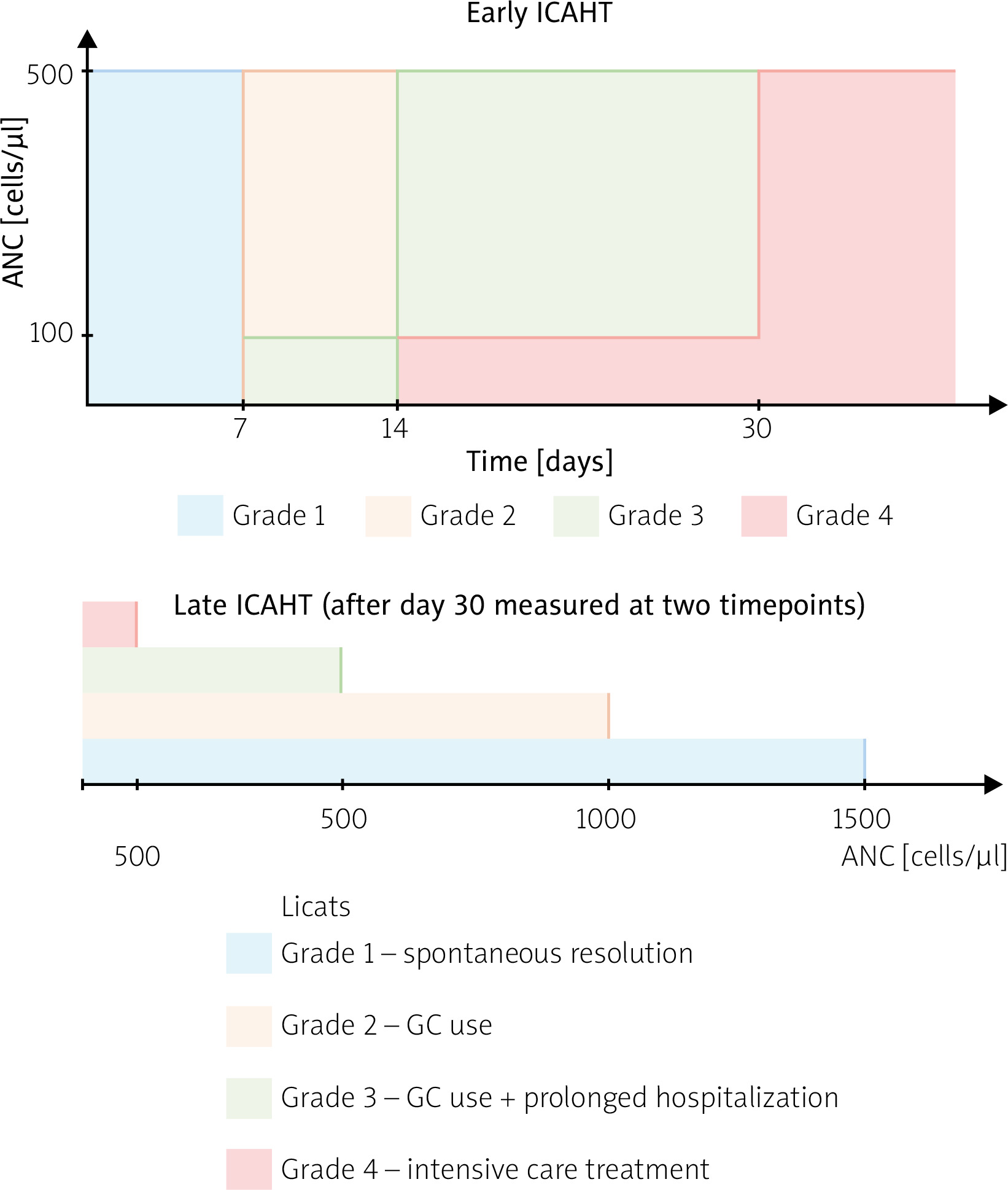
Recent scientific reports have focused on evaluating the efficacy and safety of CD19 CAR-T therapy in the treatment of SSc. Grade 1 CRS was observed in 12 of 21 patients (57%), while grade 2 CRS occurred in 2 of 21 patients (10%) who received CAR-T therapy. No cases of CRS above grade 2 were reported [7–9, 14, 15]. In the CASTLE study, the median time to onset of CRS was 1 day, and the median duration of CRS was 2 days [15]. In most cases of CAR-T therapy use in SSc, conservative treatment of grade 1 CRS proved sufficient. Müller et al. [15] administered tocilizumab to 5 of 9 patients (56%) and GCs to 1 patient. Current scientific reports on SSc suggest a favorable early neurological profile for ICANS treatment in this patient cohort. This assertion is bolstered by the absence of clinically significant ICANS among SSc patients who underwent the treatment model presented [7–9, 14, 15]. However, the current sample size of treated patients is insufficient to rule out the possibility of rare or delayed neurotoxicity. Compared with hematologic malignancies, SSc appears to be associated with a lower incidence and reduced severity of treatment-related toxicity. This phenomenon can be attributed to the lower degree of “burden” on target cells in patients with autoimmune diseases [17, 18, 22, 23]. Criteria for bone marrow suppression associated with lymphodepletion therapy are shown in Figure 2.
Other adverse reactions
Potential AEs of CAR-T therapy in SSc, in addition to the previously mentioned CRS and ICANS, encompass prolonged cytopenia [24]. Müller et al. [15] attempted to standardize criteria for reporting myelotoxicity associated with lymphodepletion therapy using immune effector cell-associated hematotoxicity (ICAHT) and local immune effector cell-associated toxicity (LICATS). Late ICATS occurred in 6/9 (66%) patients, early ICAHT in 3/9 (33%), and late ICAHT in 1/9 (11%) [15]. These criteria require wider dissemination and further validation, but they may prove useful for reporting this type of AE. Dahunsi et al. [25] posited that the administration of granulocyte-colony stimulating factor and blood transfusions is a recommended course of action. Furthermore, in cases of refractory thrombocytopenia, thrombopoietin agonists are recommended. As demonstrated in the existing body of research, delayed infections are among the most significant late complications, necessitating antiviral prophylaxis and immunoglobulins. The pathomechanism of this phenomenon has been determined to result from prolonged B-cell aplasia and hypogammaglobulinemia. In addition, a potential AE associated with the treatment is the risk of secondary malignancies, particularly myeloid malignancies, although the incidence remains low [26]. Pecher et al. [9] reported a case of a patient who developed fatal secondary hemophagocytic lymphohistiocytosis, which was probably caused by a herpes simplex virus infection.
Having considered both the clinical efficacy outcomes and the distinct toxicity profiles of CD19-targeted CAR-T cell therapy, a comprehensive synthesis of the advantages and disadvantages of this emerging modality compared to the current gold standard, autologous HSCT, is provided in Table III [27, 28].
Table III
Advantages and disadvantages of CAR-T vs. HSCT therapies in SSc
[i] The data on CAR-T therapy are taken from studies included in our review, whereas the data on HSCT are taken from systematic reviews [27, 28]. Auto-HSCT – autologous hematopoietic stem cell transplantation, CAR-T – chimeric antigen receptor T-cells, CRS – cytokine release syndrome, CYC – cyclophosphamide, FLU – fludarabine, HSCT – hematopoietic stem cell transplantation, ICANS – immune effector cell-associated neurotoxicity syndrome, MAC – myeloablative conditioning, mRSS – modified Rodnan skin score, OS – overall survival, SPM – second primary malignancy, SSc – systemic sclerosis, TBI – total body irradiation, TRM – treatment-related mortality.
Limitations of chimeric antigen receptor T-cell therapy in systemic sclerosis
Notwithstanding early encouraging observations of CAR-T therapy in the treatment of autoimmune diseases, the therapeutic modality is also associated with numerous significant limitations. Primarily, these limitations pertain to small patient cohorts and limited databases, with many conclusions based on analyses of individual cases. The consequence is difficulty in monitoring the therapy’s effectiveness in a wider group of recipients, which creates challenges in future assessments of its effectiveness and safety [29]. Another important aspect is the lack of available randomized studies. Contemporary scientific reports indicate that the limited duration of observation constitutes a significant challenge. This limitation adversely impacts the capacity to ascertain data about the durability of remission and to estimate the likelihood of subsequent AEs [30]. A further challenge lies in the divergence of protocols, the absence of a clearly delineated standard of care, and the heterogeneity of methodologies across studies, rendering comparisons of results across centers a formidable task. This circumstance creates considerable challenges in identifying the key elements that contribute to achieving the intended therapeutic outcome [30, 31]. This treatment method necessitates substantial financial expenditures and the establishment of specialized infrastructure, factors that consequently restrict its accessibility [32].
Future directions for the development of chimeric antigen receptor T-cell therapy in systemic sclerosis
Recent perspectives on the treatment of SSc using CAR-T cell therapy emphasize the need for prospective studies with standardized protocols and long-term monitoring. This is significant because such observations facilitate analysis of remission in terms of durability and enable evaluation of a comprehensive safety profile [14, 29, 33, 34]. The authors likewise underscore the need to leverage inflammatory response markers to facilitate optimal patient selection. Moreover, this approach enables us to determine whether early intervention in patients with an aggressive phenotype has a lasting impact on disease progression [14, 29, 33]. A significant avenue for advancement lies in implementing readily available allogeneic formulations that reduce qualification waiting periods and minimize costs. Additional genetic manipulations remain crucial to ensure the safety and efficacy of these “off-the-shelf products.” These include the use of CRISPR/Cas9 technology. Gene editing primarily leads to the elimination of endogenous T-cell receptor (TCR) expression. Consequently, allogeneic CAR-T cells become undetectable to the immune system, reducing the risk of graft-versus-host disease (GvHD). To prevent the immune system from rejecting therapeutic cells, molecules such as human leukocyte antigens (HLA) or CD52 are eliminated in most cases. Another significant aspect, apart from gene editing, is the appropriate selection of the cell source, which can influence the incidence of GvHD. For example, using regulatory T cells (Tregs) or NK cells as the basis for CAR modification may reduce the risk of GvHD compared to conventional T cells. CARs can be synthesized on the surface of conventional T cells, Tregs, NK cells, or macrophages, thereby enabling these cells to acquire distinct functional characteristics. In addition, important challenges remain in CAR-based allogeneic therapy, and solutions are still pending. Nonetheless, several preparations using universal platforms have entered clinical trials. A case study demonstrating the feasibility of achieving deep remission and regression of fibrotic changes while ensuring safety is provided by an analysis employing TyU19 (which uses CRISPR/Cas9 to achieve these exact gene knockouts) in 2 patients with dSSc. This finding reinforces the prospect of future observations regarding the universality of products obtained using CRISPR [8, 33–35]. Concurrently, alternative methods of deep B-cell depletion are being promoted; these may either compete with or coexist with standard CD19-CAR-T. Numerous scientific reports on rheumatic diseases and SSc underscore the distinctive role of bispecific antibodies, exemplified by blinatumomab. These antibodies effectively facilitate the targeting of autologous T cells against CD19+, eliminating the need for viral vectors and enhanced conditioning [30, 33, 36]. The future of CAR-T therapy for SSc holds enormous potential, but it remains in its early stages of development. Several trials are underway to further assess the efficacy and safety of CAR-T cells in SSc.
An overview of CAR-T therapy in the treatment of SSc is presented in Supplementary Table I.
Conclusions
CD19-targeted CAR-T therapy currently represents an early proof of concept in patients with treatment-refractory SSc, with promising initial clinical responses, including reductions in skin fibrosis and stabilization of organ involvement. The early safety profile appears favorable, characterized primarily by low-grade CRS and a notable absence of severe neurotoxicity, which compares positively to procedures such as HSCT. Despite these promising results, the available evidence is largely based on individual case reports and heterogeneous groups, with limited observation periods. Consequently, the long-term safety and durability of remission remain unproven. Further validation through long-term randomized controlled trials is therefore essential. Advances in allogeneic “off-the-shelf” products and bispecific antibodies may help to standardize and expand clinical access to this new therapeutic strategy.