Introduction
Systemic lupus erythematosus (SLE) is a chronic autoimmune and inflammatory disease with multiple organ manifestations. Serous membranes are affected in about 10–50% of SLE patients, mostly pleura and pericardium [1]. In SLE patients, risk factors for serositis are defined as male sex, level of anti-double stranded-DNA (anti-ds-DNA) antibody, febrility, active SLE, D-dimer, low complement, elevated sedimentation rate, lupus nephritis, interstitial lung disease, pulmonary arterial hypertension, and cytopenias [1–3]. Serositis association with anti-Smith protein (anti-Sm), anti-ribonucleoprotein (anti-Rnp) and antiphospholipid autoantibodies (APL) is documented [4]. Systemic lupus erythematosus in patients older than 50–65 years or elderly onset (EO) SLE is regarded by some authors as a different disease phenotype with higher serositis prevalence [5–7]. One EO cohort suggests a more indolent course of SLE but a high burden of comorbidities in such patients [4]. Peritoneum is rarely inflamed, and very few patients with SLE have peritonitis complicated with ascites. Serum tumor marker CA-125 may be independently associated with serositis [6, 8]. When SLE serositis and pleural, pericardial, or peritoneal effusions are associated with false positive ovarian tumor markers (TM) and with no underlying malignancy, this condition is referred to as pseudo-pseudo-Meigs’ syndrome (PPM) or Tjalma syndrome (TS) after the physician who first described it in 2006 [9]. The therapeutic approach to TS is not standardized. Usually induction therapy consists of short duration methylprednisolone pulse therapy and a glucocorticosteroid (GC) tapering scheme. Remission maintenance is with various immunosuppressants including rituximab [10].
Material and methods
A literature review was performed to identify previously reported cases and studies related to TS. Articles published between 2008 and 2025 were searched using several electronic databases, including PubMed/MEDLINE, Web of Science, and Google Scholar. Additional relevant articles were identified through reference lists of selected publications. The search was conducted using combinations of the following key words: “Tjalma syndrome”, “pseudo-pseudo Meigs syndrome”, “systemic lupus erythematosus”, “ascites”, and “pleural effusions.” Boolean operators (AND/OR) were used to refine the search. Articles were included if they: reported cases or case series describing TS or PPM syndrome; were published between 2008 and 2025; were available in English and contained sufficient clinical information for analysis. Articles were excluded if they: were duplicate reports; did not describe clinical cases of TS; or lacked relevant clinical data. Data extracted from the selected articles included year of publication, patient demographics, clinical presentation, associated conditions, treatment modalities, and outcomes. The findings from identified studies were summarized and presented in a tabular format.
The procedures followed in this study were in accordance with the ethical standards of the Helsinki Declaration of 1964, as revised in 2013. The patient has provided written informed consent for publication of the case details.
Case description
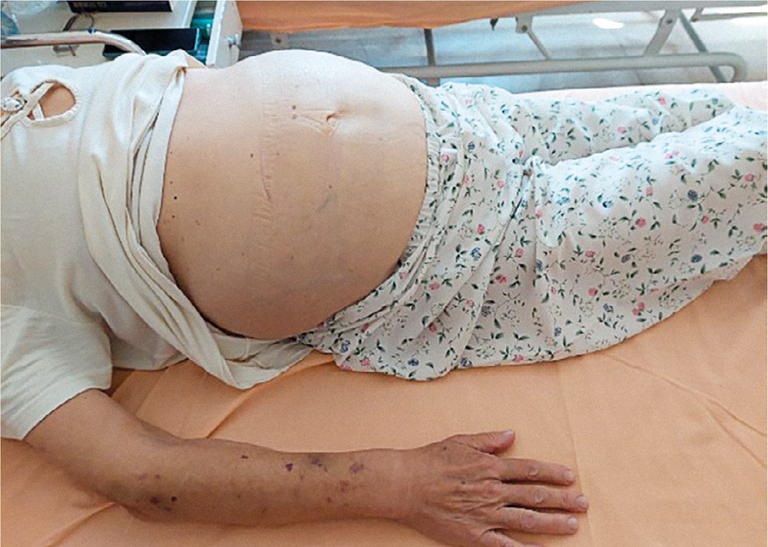
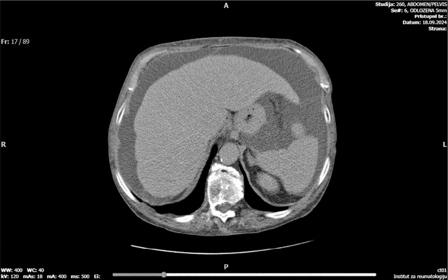
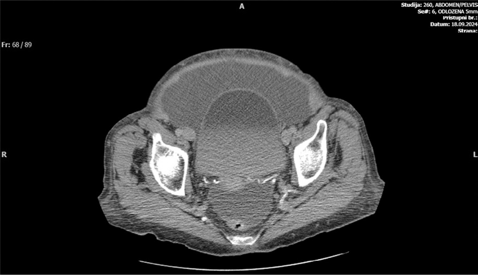
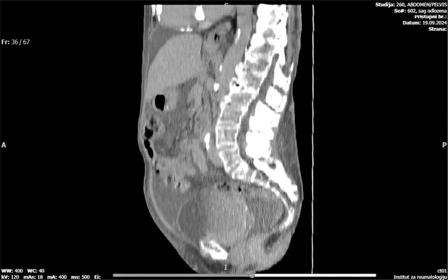
The 67-year-old female patient with a 5-year history of SLE was admitted to the hospital in September 2024, because of large, slowly growing ascites (Figure 1). Systemic lupus erythematosus started in 2019. and was diagnosed based on constitutional symptoms, bilateral pleural, pericardial and peritoneal effusions, antinuclear antibodies positivity, typical skin changes, myalgias and cytopenias (the patient fulfilled 2019 Alliance of Associations for Rheumatology [EULAR]/American College of Rheumatology [ACR] criteria for SLE: ANA positivity as the entry criterion and a total score of 17 points across the constitutional, hematological, mucocutaneous, and serosal domains. The patient was treated with GCs and antimalarials from the onset of her disease. On admission, her comorbidities, important for present ascites etiology, were all well controlled: cardiomyopathy, diabetes with chronic renal disease, and hypothyroidism. Physical findings revealed: systolic heart murmur, iatrogenic Cushing’s syndrome (typical obesity distribution and parchment-like skin) and lower legs’ chronic venous dermatitis. Laboratory findings showed: C-reactive protein (CRP) 14.3, mild pancytopenia, and stable chronic renal disease parameters. The last SLE flare was in October 2023, when abdominal and pelvic ultrasound detected perihepatic, perisplenic, bilateral paracolic, and pelvic ascites treated with GCs and diuretics. Transthoracic echocardiography was urgently performed and excluded visible endocarditis and decompensated cardiomyopathy. Elevated mean right atrial pressure of 35 mmHg was detected, similar to the echocardiography finding from October 2023. The patient received prednisone (20 mg once daily) and continued hydroxychloroquine (HCQ; 200 mg once daily). Ascites was treated with larger doses of furosemide, two therapeutic paracenteses with 8 liters of fluid evacuated, and parenteral antibiotics. The patient had significantly high ovarian tumor markers levels in serum and ascites (CA-125, HE4 and Roma index) (Tables I and II). Multidetector computed tomography (MDCT) of the abdomen and pelvis was significant for large ascites (Figures 2–4), along with signs of possible Budd-Chiari syndrome, and excluded malignancy. The patient was started on 2 × 0.4 ml subcutaneous fraxiparine but developed significant thrombocytopenia the next day. Low molecular weight heparin was stopped, and apixaban 2.5 mg twice daily was introduced. Weak positive anti-HIT antibodies were detected. During hospitalization the patient developed COVID-19 infection with no signs of pneumonia, but with worsening pancytopenia. Multidetector computed tomography and magnetic resonance imaging excluded thrombotic hepatic disease, and no tumor was found. The patient received three consecutive days (250 mg daily) of methylprednisolone pulse therapy followed by prednisone 20 mg twice daily (with tapering scheme), HCQ 200 mg once daily, and azathioprine 50 mg once daily with cautious dose escalation due to cytopenias.
Table I
Immunological markers
[i] AB – antibodies, ACLA – anticardiolipin antibodies, ACPA – anti-citrullinated peptide antibodies, AMA – anti-mitochondrial antibodies, ANA – antinuclear antibodies, anti-β2GPI – anti-β2-glycoprotein I antibodies, APA – antiparietal antibodies, ASMA – anti-smooth muscle antibodies, C3 and C4 – complement components, RF – rheumatoid factor.
Table II
Longitudinal serum, urine, and ascites laboratory values monitoring (selected abnormal values are bolded)
[i] C-reactive protein: < 5 mg/l, leukocyte absolute count: 3.4–9.7 × 10⁹/l, CA-125 – up to 35 U/ml, lymphocyte absolute count: 1.2–3.4 × 10⁹/l, hemoglobin ref.: 119–157 g/l, platelets ref.: 158–424 × 109/l, urea: 2.8–7.2 mmol/l, creatinine 58–96 µmol/l, albumin ref.: 35–53 g/l, γ-glutamyl transferase ref.: 9–39 U/l, , human epididymis protein 4 – before menopause ≤ 73 pmol/l, after menopause ≤ 93 pmol/l, procalcitonin ref.: > 2.0 ng/l high risk of systemic infection.
[ii] AARB – acid-alcohol resistant Bacilli, Alb – albumin, BNP – blood natriuretic peptide, BUN – blood urea nitrogen, CA – 125-cancer antigen, Cre – creatinine, CRP – C-reactive protein, DAT – Coombs test, FOBT – fecal occult blood test, Glc – glucose, GGT – γ-glutamyl-transferase, Hapt – haptoglobin, HE4 – human epididymis protein 4, Hgb – hemoglobin, LDH – lactate dehydrogenase, LYMPH – lymphocytes, PCT – procalcitonin, PTL – platelets, TnT – troponin, WBC – white blood cells.
Discussion
Systemic lupus erythematosus is an autoimmune inflammatory disease that predominantly affects women during the reproductive period of life. A late-onset phenotype also exists and represents a diagnostic and therapeutic challenge due to its clinical presentation and the presence of comorbidities in elderly patients. Inflammation of the epithelium lining the serous cavities is considered a severe manifestation of the disease and requires careful differential diagnosis as well as aggressive anti-inflammatory treatment.
The differential diagnosis of pericardial, pleural, and/or peritoneal effusions in elderly patients burdened with cardiovascular and other comorbidities includes heart failure, hypothyroidism, liver insufficiency, infections such as tuberculosis, and paraneoplastic manifestations. Determination of tumor marker levels in the presence of serositis may yield false-positive results; therefore, ovarian tumor markers such as CA-125 and HE4 may also be elevated in male patients with serositis.
Serositis in lupus is considered a severe systemic manifestation that requires intensive anti-inflammatory treatment with pulse doses of GCs and appropriate maintenance therapy. However, in elderly patients such therapy may worsen the overall internal clinical status. When a paraneoplastic syndrome or malignant disease is suspected, necessary intensive treatment may be delayed due to concerns about possible deterioration of the underlying condition or progression of the primary disease.
In our patient, we firstly ruled out primary and metastatic malignant disease of the ovaries by the gynecologist’s directed diagnostic imaging: ultrasound and MDCT. After effusion puncture, no malignant cells were detected. We did not use positron emission tomography scan because of the high probability of indeterminate results.
All reported cases included in this article describe female patients [11–31]. Our patient had LO SLE diagnosed in 2019 with serositis as the first disease manifestation. Due to her comorbidities, namely chronic non-lupus-related renal disease and cardiomyopathy, the patient was thoroughly monitored by a nephrologist and cardiologist, and effusions were not associated with these systemic causes. In the past, patient’s SLE serositis flares responded well to GC therapy, but no adequate GC-sparing immunosuppressive was introduced, except for immunomodulatory antimalarial. Patient’s serositis episodes were not associated with worsening of SLE immunological markers: anti-ds-DNA levels and complement levels were normal. No concomitant or overlap autoimmune disease was diagnosed, as scleroderma and myositis antibodies profiles were normal and mixed connective tissue disease was excluded.
Age of onset of TS in case reports and case series from Table III ranges from 14 years old to 82 years old, but most patients are younger than 50 years old [11–31]. Most patients have TS as an initial SLE manifestation of rheumatic disease [11–31]. Our patient had SLE for 4 years, before she was diagnosed with ascites. Patient’s latest SLE flare was in October 2023, with slowly growing ascites at first resistant to GCs and diuretics. Other SLE manifestations in our patient are intermittent cytopenias. Authors have reported cases with SLE nephritis [12, 13, 26] and arthritis [14, 25], but our patient has no history of either condition. Most authors have reported TS with ascites and unilateral or bilateral pleural effusions [11–31]. The most common therapy for remission induction is high-dose intravenous methylprednisolone, following different prednisone regimens and immunosuppressive agent introduction [11–20, 22–26, 29, 30]. Our patient’s SLE-related serositis and both pleural and peritoneal effusions were treated with methylprednisolone pulses, then oral prednisone with a tapering scheme and azathioprine as an immunosuppressive. During a few monthly follow-ups, the patient had no significant relapses.
Table III
Case reports and case series with TS
| Case | Year | Sex | Age | Disease duration | SCTD | Serositis | Treatment |
|---|---|---|---|---|---|---|---|
| [11] | 2024 | F | 27 | 0 | Bullous SLE | Pleural uni | MP, HCQ |
| [12] | 2024 | F | 33 | ND | SLE nephritis | Pleural bil Ascites | MP, HCQ, MMF |
| [13] | 2024 | F | 28 | 0 | SLE | Pleural bil Ascites | MP, PRN, MMF, telitacicept |
| F | 37 | 0 | Sjögren’s disease SLE nephritis | Pleural bil Ascites Pericardial | MP, PRN, MMF, telitacicept | ||
| F | 50 | 10 | Sjögren’s disease SLE | Pleural bil Ascites Pericardial | MP, CyP | ||
| [14] | 2023 | F | 32 | 8 | SLE Arthritis | Pleural bil | MP, PRN, HCQ, MTX |
| [15] | 2023 | F | 33 | ND | SLE | Pleural bil Ascites | MP, MMF |
| [16] | 2023 | F | 74 | 7 | SLE | Pleural bil Ascites | MP, HCQ, MMF |
| [17] | 2022 | F | 82 | 0 | SLE | Pleural Ascites | GCS, HCQ |
| [18] | 2022 | F | 23 | 0 | SLE Cystitis | Ascites | PRN, HCQ |
| [19] | 2022 | F | 41 | 0 | SLE | Pleural uni Ascites | MP, PRN, HCQ, MMF |
| [20] | 2022 | F | 48 | 0 | SLE | Pleural Ascites | MP, PRN, CYP |
| [21] | 2022 | F | ND | ND | SLE Protein-losing enteropathy | ND | ND |
| [22] | 2021 | F | 23 | ND | SLE | Pleural Ascites | MP, PRN, HCQ, AZA |
| [23] | 2019 | F | 44 | 0 | SLE Cytopenias | Pleural | MP, HCQ, LEF |
| [24] | 2019 | F | 24 | 4 | SLE | Pleural bil Ascites | MP, MMF |
| [25] | 2019 | F | 44 | ND | SLE Arthritis | Pleural bil Ascites | MP, PRN, AZA |
| [26] | 2019 | F | 22 | ND | SLE APS Nephritis | Pleural Ascites | MP, PRN, HCQ |
| [27] | 2019 | F | 14 | ND | ND | Pleural Ascites | ND |
| [28] | 2016 | ND | ND | ND | MCTD | Pleural Ascites | ND |
| [29] | 2016 | F | 40 | 0 | SLE | Ascites | MP |
| [30] | 2014 | F | 22 | ND | SLE | Ascites | Leiomyoma |
| [31] | 2008 | F | 38 | 0 | SLE | Pleural Ascites | ND |
[i] APS – antiphospholipid syndrome, AZA – azathioprine, CyP – cyclophosphamide, HCQ – hydroxychloroquine, LEF – leflunomide, MP – methylprednisolone, MCTD – mixed connective tissue disease, MMF – mycophenolate mofetil, MTX – methotrexate, ND – no data, Pleural uni – unilateral pleural, Pleural bil – bilateral pleural, PRN – prednisone, SCTD – systemic connective tissue disease.
Literature analysis and presented case studies are aimed at raising awareness of this rare manifestation of SLE, particularly in EO SLE with high suspicion of paraneoplastic syndrome. A thorough diagnostic approach and carefully tailored immunosuppressive therapy are of great importance.