Introduction
Osteoporosis represents one of the most prevalent skeletal disorders worldwide, characterized by reduced bone mineral density and disruption of bone microarchitecture. These pathological changes significantly increase the risk of low-trauma, or fragility, fractures, which are associated with considerable morbidity, decreased quality of life, and elevated mortality rates. Fragility fractures reflect compromised bone strength and may be either clinically silent or manifest with pain and structural deformity [1].
Given its widespread occurrence, osteoporosis represents a significant global public health challenge, with approximately one in 2 women and one in 5 men over the age of 50 projected to experience a fragility fracture during their remaining lifetime [2]. As the disease typically develops silently, fractures are commonly the first indication of its presence. This emphasizes the urgent need for improved insight into its underlying biology to support earlier diagnosis and more effective treatment strategies [3].
According to the World Health Organization (WHO) criteria established in 1994 and still in use, osteoporosis is defined as a bone mineral density (BMD) value of at least 2.5 standard deviations below the mean peak BMD of young, healthy women, corresponding to a T-score of less than –2.5 [4]. Densitometry (dual-energy X-ray absorptiometry – DXA) remains the gold standard in the diagnosis of osteoporosis, although it undoubtedly has several limitations: it may overestimate bone density in individuals with spinal or hip degenerative changes, and artefacts from osteophytes, prior surgery, or vascular calcifications can distort spine BMD results. Additionally, the technique lacks information about bone microarchitecture [5].
In addition to DXA, bone turnover markers, including procollagen type I N-propeptide (PINP), bone-specific alkaline phosphatase (BALP), β-isomerized C-terminal telopeptide of type I collagen (β-CTX-I), and tartrate-resistant acid phosphatase 5b (TRACP5b), constitute an additional group of biomarkers relevant to osteoporosis assessment [6]. Serum PINP and plasma β-CTX-I are commonly used reference markers for monitoring response to anti-osteoporotic therapy, while BALP and TRACP5b may be particularly useful in patients with chronic kidney disease due to their limited dependence on renal function. Despite this, it is worth remembering that bone turnover markers are characterised by high biological and analytical variability and may fluctuate, for example, due to recent food intake.
Owing to the limitations of the currently used methods, there is a pressing need for reliable and widely accessible biomarkers for the early detection and monitoring of this condition.
Since osteoporosis results from dysregulated bone remodelling, characterized by bone resorption exceeding bone formation, recent research has increasingly focused on the role of microRNAs (miRNAs) in modulating this imbalance [3]. Currently, miRNAs are being investigated for use in both the diagnosis and treatment of osteoporosis. Additionally, there is an attempt to commercialise the solutions developed so far.
MicroRNA characterisation
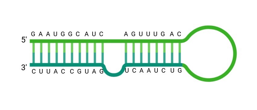
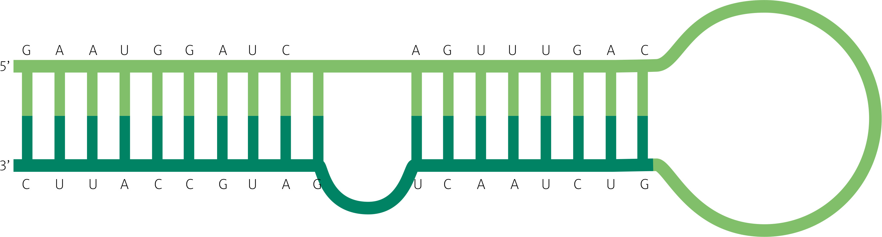
MicroRNAs constitute a class of endogenous, small non-coding RNA molecules typically comprising 20–24 nucleotides that play a pivotal role in post-transcriptional gene regulation [7–9]. The schematic structure of a pre-miRNA that undergoes maturation to generate a functional miRNA that interacts with messenger RNA (mRNA) is presented in Figure 1. The human genome encodes approximately 2,000 miRNAs [10]. It is estimated that microRNAs may regulate up to 60% of human protein-coding genes. Their mechanism of action involves binding to the 3´ untranslated regions (3´UTR) of target mRNAs, leading to transcript degradation or suppression of translation [3, 11]. The regulatory network of miRNAs is highly intricate, as individual miRNAs can influence multiple gene targets, and conversely, a single mRNA can be modulated by several different miRNAs. They act primarily by inhibiting translation or promoting the degradation of target mRNAs. Dysregulated miRNAs can lead to changes in gene expression that impact many aspects of health and contribute to a variety of human diseases. Due to their remarkable stability in biological fluids and being measurable using well-established quantification methods, circulating miRNAs have gained significant attention as non-invasive biomarkers for disease diagnosis and prognosis. Aberrations in miRNA expression are increasingly implicated in the pathogenesis of a wide range of human disorders, such as cancer, metabolic diseases, and musculoskeletal conditions including osteoporosis [3]. Moreover, miRNA-based therapeutic strategies represent a promising frontier in translational medicine. Since their initial discovery, an expanding range of miRNAs has been identified, accompanied by rapid advances in elucidating their functional roles and potential clinical applications.
Fig. 1
Structure of a precursor microRNA (pre-miRNA) which undergoes maturation to generate a functional miRNA that interacts with mRNA. Created with biorender.com.
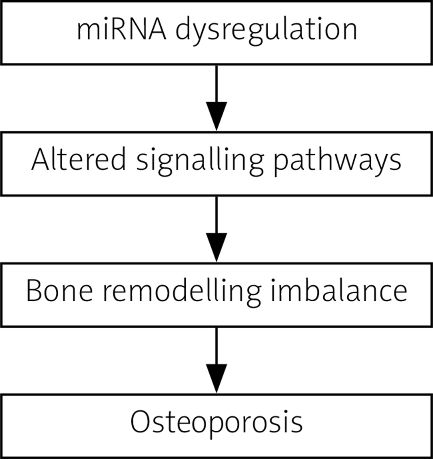
Accumulating evidence also suggests that epigenetic mechanisms play a significant role in the pathogenesis of osteoporosis.
The influence of miRNA in the pathomechanism of osteoporosis is presented in Figure 2.
The role of microRNAs in osteoclastogenesis and bone resorption
Interest in the role of miRNAs in osteoporosis has been steadily increasing for over a decade. Aberrant expression of specific osteoclast-associated miRNAs has been implicated in the pathogenesis of osteoporosis through the promotion of excessive bone resorption. Some of the earliest studies published in this field investigated the function of miR-133a in osteoclastogenesis [12]. The objective of one of the first studies, conducted by Wang et al. [12], was to identify key miRNAs in circulating human monocytes that are linked to differing bone mineral density (BMD) profiles among postmenopausal Caucasian women. Notably, both microarray and quantitative reverse transcription polymerase chain reaction (qRT-PCR) analyses revealed a significant elevation of miR-133a expression in individuals with low BMD. Subsequent authors have confirmed the role of miR-133a in osteoclastogenesis [13, 14]. It was demonstrated that miR-133a was overexpressed in postmenopausal women with low bone mineral density.
Efforts were also made to elucidate the mechanism of action of miR-133a in the context of osteoporosis. Research by Wang et al. [15, 16] demonstrated that miR-133a overexpression inhibited the osteogenic differentiation of bone marrow-derived mesenchymal stem cells (BMSCs). In contrast, silencing miR-133a exerted beneficial effects on mesenchymal stem cells exposed to glucocorticoids and mitigated bone loss in animal models of glucocorticoid-induced osteoporosis. These effects were mediated via the mitogen-activated protein kinases/extracellular signal-regulated kinase (MAPK/ERK) signalling pathway through the targeting of fibroblast growth factor receptor 1. Similarly, Li et al. [14] reported that suppression of miR-133a influenced serum levels of osteoclastogenesis-related markers, enhanced lumbar spine bone mineral density, and improved bone histomorphometry in ovariectomized rats. On the other hand, Zhou et al. [17] proposed that miR-133a expression in osteoblasts helped to preserve bone structure and mechanical strength in mice subjected to mechanical unloading, thus reducing bone loss. Despite some differences in experimental approaches, the regulatory direction of miR-133a across these studies was generally consistent, indicating its potential as a biomarker for osteoporosis diagnosis.
Another miRNA molecule associated both with promoting osteoclastogenesis and inhibiting osteogenesis is miR-214-3p. Consistent findings have indicated that miR-214-3p is upregulated in individuals with osteoporosis [18–21]. It exerts multifactorial and complex regulatory effects. MiR-214-3p has been reported to suppress osteogenic differentiation through multiple mechanisms of action, one of which involves the inhibition of osterix, which is required for the differentiation of preosteoblasts into functional osteoblasts [22]. The study conducted by Mohamad et al. [18] explored the potential involvement of osterix and miR-214 in primary osteoporosis by analysing their expression levels in bone samples obtained from individuals with and without osteoporosis, and by examining their relationships with each other as well as with clinical and laboratory parameters. The findings revealed that patients with osteoporosis undergoing joint replacement surgery exhibited significantly elevated levels of miR-214 compared to non-osteoporotic individuals. Consequently, osterix expression showed an inverse pattern, with markedly lower levels observed in osteoporotic patients [18]. Also, in the research performed by Shi et al. [20] miR-214-3p expression was inversely correlated with osterix expression. This is not the only mechanism of action of miR-214-3p, which has also been shown to inhibit osteogenic differentiation through the suppression of key regulatory factors such as ATF4 (a gene encoding one of the main transcription factors required for osteoblast function), and fibroblast growth factor receptor 1 [23–25]. Among the numerous mechanisms by which miRNA-214-3p influences bone metabolism, one notable pathway involves the promotion of osteoclastogenesis through targeting phosphatase and tensin homolog (PTEN), which results in promoting osteoclastogenesis [23].
MicroRNA-mediated regulation of osteogenesis
An additional miRNA implicated in the regulation of bone metabolism is miR-125b. The expression level of miR-125b has been reported to be upregulated in patients with osteoporosis, and it is considered to be responsible for the development of postmenopausal osteoporosis. Among the miRNAs expressed in osteoblasts, miR-125b has been identified as being selectively incorporated into matrix vesicles and deposited into the bone matrix. During bone resorption, it is subsequently released into the bone marrow microenvironment, where it contributes to the suppression of osteoclastogenesis. This regulatory effect is mediated through the inhibition of PR domain zinc finger protein 1 (PRDM1) expression – a transcriptional repressor that negatively modulates factors involved in the inhibition of osteoclast formation [26].
Further analysis demonstrated that miR-125b negatively regulates the expression of osteogenic marker genes throughout this differentiation process, indicating that its downregulation is essential for promoting osteogenesis [27]. Bioinformatic predictions identified bone morphogenetic protein receptor type 1B (BMPR1b) as a potential target of miR-125b. This interaction was confirmed through a dual-luciferase reporter assay, which revealed that miR-125b directly binds to the 3´-untranslated region (3´UTR) of the BMPR1b mRNA. Silencing BMPR1b significantly impaired the osteogenic differentiation capacity of human BMSCs.
Studies investigating the clinical context of miR-125b regulatory mechanisms conducted in postmenopausal women have demonstrated significant upregulation of miR-125b expression in both blood and bone tissue of individuals diagnosed with osteoporosis [28, 29]. Interestingly, the results of one study are not consistent with those of the others [30]. The research conducted by Chen et al. [30] revealed decreased expression of miR-125b-5p in the osteoporosis group, suggesting downregulation rather than upregulation. This discrepancy in the results may be attributed to differences in patient characteristics, as the osteoporotic individuals in that study had not experienced recent fractures within the preceding two months. It is possible that circulating miRNA profiles vary between osteoporotic patients with fractures, those without fractures, and non-osteoporotic controls.
Complex regulatory role of miR-21-5p in osteoporosis pathophysiology
Despite being one of the most thoroughly investigated microRNAs, the functional contribution of miR-21-5p to osteoporosis pathogenesis appears to be complex and not yet fully defined. The aberrant regulation of miR-21-5p in osteoporosis has emerged as a focal point of recent scientific inquiry [31]. The regulatory pattern of miR-21-5p in osteoporosis appears to be conflicting – while some studies have reported elevated expression [32–35], others have observed downregulation [13, 35]. Such variability may reflect the multifaceted role of miR-21-5p in modulating both bone formation and resorption processes through diverse signalling mechanisms, i.e. by influencing both osteoblast and osteoclast differentiation through various molecular pathways. It has been shown to promote osteogenesis, yet its role in osteoclastogenesis remains complex and partly contradictory. Some studies demonstrate that miR-21-5p enhances osteoclast formation, while others suggest that it suppresses osteoclast differentiation.
Despite these inconsistencies, it is clear that miR-21-5b plays a role in bone metabolism. As of now, its use as a diagnostic biomarker is limited, and further research is needed to elucidate the reason behind the discrepancies between the studies.
Additional microRNAs with potential as osteoporosis biomarkers
The miRNAs described above are not the only ones investigated in the context of osteoporosis; however, they exemplify the current direction of research in this field. Additional studies have also examined the potential roles of miR-483-5p [36, 37], miR-497-5p [38, 39], and miR-422 [40] in the pathogenesis and progression of this condition. Interestingly, only one microRNA molecule, miR-497-5p, has been found to be downregulated, distinguishing it from the majority of miRNAs investigated in the context of osteoporosis. The most relevant miRNAs involved in the pathophysiology of osteoporosis are summarized in Table I.
Advances in microRNA-based therapies for bone regeneration
In parallel with research on the potential use of miRNAs in the diagnosis of osteoporosis, efforts have also begun to explore their therapeutic applications in the treatment of this disease. In general, two principal strategies are employed in miRNA-based therapies, depending on the nature of miRNA dysregulation [41]. miRNA replacement therapy is used when a pro-osteogenic miRNA is downregulated. In this case, synthetic miRNA mimics are introduced to restore physiological function by targeting the same mRNAs as endogenous miRNAs, potentially enhancing osteoblast activity – for instance, using miR-29b mimics to stimulate bone formation [42]. Conversely, miRNA inhibition is applied when a deleterious miRNA, such as a pro-resorptive or anti-osteogenic one, is pathologically overexpressed. Synthetic antisense oligonucleotides (e.g., antagomiRs – synthetic, chemically modified single-stranded RNA molecules designed to specifically inhibit the activity of a miRNA) bind to and neutralize these miRNAs, preventing their interaction with target transcripts. An example is the inhibition of miR-214 to promote osteogenesis.
Although miRNA-based therapies hold substantial promise, their clinical application remains limited by challenges in efficient in vivo delivery [43]. Naked RNA molecules are unstable in circulation due to nuclease degradation and are poorly internalized by cells because of their size and negative charge. Therefore, creating effective delivery systems is key for using miRNAs as therapies. Researchers are exploring both viral carriers, such as adeno-associated viruses, and non-viral alternatives [41]. While viral vectors offer high transduction efficiency and sustained expression, their use is restricted by immunogenicity and safety concerns. Non-viral systems –including lipid nanoparticles, polymer-based carriers, and exosome-based delivery – offer safer alternatives with potential for clinical translation.
In research conducted so far to overcome the limitations of current well-established osteoporosis therapies, a gene therapy approach was developed that targets two key miRNAs – miR-214-3p and miR-34a-5p – simultaneously promoting osteogenesis while suppressing osteoclastogenesis [44]. This strategy employed bone-targeting recombinant adeno-associated viral (rAAV) vectors to systemically deliver gene modulators directly to skeletal tissue. In murine models, overexpression of miR-214-3p or suppression of miR-34a-5p induced osteoporotic-like bone loss, while inhibition of miR-214-3p or upregulation of miR-34a-5p reversed bone degradation in both postmenopausal and senile osteoporosis. These effects were achieved by increasing osteoblast activity and reducing osteoclast function. Importantly, the therapeutic intervention did not elicit pathological changes in non-skeletal tissues.
Translation into clinical testing
Unlike intracellular RNA molecules, extracellular miRNAs display remarkable stability – they withstand RNase activity and remain intact even under extreme conditions such as boiling, repeated freeze–thaw cycles, and highly alkaline (pH 13) or acidic (pH 1) environments [45, 46]. However, there are a few technical challenges related to the methods used for analysing circulating miRNAs: miRNA concentration in plasma/serum is significantly lower than in cells or tissues; miRNA composition in biofluids can depend on sample collection procedures; and the presence of enzyme inhibitors or contaminating cells can influence miRNA data. That is why assays must be validated for their capability to detect and quantify extracellular miRNAs, and pre-analytical procedures should be performed correctly [46]. Although the issue may appear complex, the first systems enabling population-level assessment of miRNA expression in the context of osteoporosis are already available [47].
Conclusions and future perspectives
Osteoporosis still poses persistent challenges in both diagnosis and treatment, and there is a need to identify novel disease biomarkers and to develop innovative therapeutic approaches. Current research suggests that miRNAs contribute to the regulation of bone metabolism. Nevertheless, evidence supporting their practical use in osteoporosis management remains limited. Their relevance for diagnostic purposes, determination of treatment thresholds, and therapeutic application has yet to be firmly established. That is why the field of miRNA research in osteoporosis is currently at a critical point. Substantial progress has been made in elucidating the biological functions of miRNAs, and their potential clinical utility is increasingly recognized.
It seems that current research should focus primarily on:
elucidating the roles of other miRNA molecules in the pathogenesis of osteoporosis,
validating previous findings on the role of already discovered miRNAs,
further work on glucocorticoid-induced osteoporosis and osteoporosis in inflammatory arthritis,
commercialisation of novel miRNA-based diagnostic tools,
evaluating the potential use of viral vectors as delivery systems for therapeutic miRNA.
Nonetheless, the translation of these molecular insights into clinical applications will remain limited unless the research focus shifts from discovery toward validation and standardization. The next major advancement in this domain is likely to be the establishment of methodological frameworks enabling the definitive validation of already-identified, high-potential candidates. Such progress will require coordinated, multidisciplinary collaboration and adherence to reproducible methods. Only through collaborative work on the complex functions of miRNAs can the research advances be translated into real-world diagnostic and therapeutic solutions for affected patients.
The use of miRNAs offers promising potential for a deeper understanding of the pathophysiology of osteoporosis, which may, in turn, enable earlier detection and more effective therapeutic interventions.